往期刊物2018

卷册: 8, 期号: 2

left

Dec

20

Dec

5

Nov

20

Nov

5

Oct

20

Oct

5

Sep

20

Sep

5

Aug

20

Aug

5

Jul

20

Jul

5

Jun

20

Jun

5

May

20

May

5

Apr

20

Apr

5

Mar

20

Mar

5

Feb

20

Feb

5

Jan

20

Jan

5

right

生物化学

鱼胆汁清除后进行酶谱和质谱蛋白质组学分析

Fish Bile Clean-up for Subsequent Zymography and Mass Spectrometry Proteomic Analyses

鱼胆汁清除后进行酶谱和质谱蛋白质组学分析

Rachel Ann Hauser-Davis Rachel Ann Hauser-Davis
7374 Views
Jan 20, 2018
Biliary excretion offers a way to analyze various contaminants in aquatic organisms, and fish bile has been used as a biomarker for environmental contamination. The use of the fish bile proteome as a tool for monitoring the impact of environmental contaminants has been recently validated. However, scarce studies in this context are available, and much remains to be investigated. In this context, this protocol describes a fast, reproducible and cheap biliary clean-up procedure for subsequent proteomic analyses, such as zymography and mass spectrometry.

细胞生物学

利用大鼠成年肝细胞生成化学诱导肝祖细胞(CLiP)

Generation of Chemically Induced Liver Progenitors (CLiPs) from Rat Adult Hepatocytes

利用大鼠成年肝细胞生成化学诱导肝祖细胞(CLiP)

TK Takeshi Katsuda
KH Kazunori Hosaka
TO Takahiro Ochiya
10894 Views
Jan 20, 2018
Primary mature hepatocytes (MHs) or their progenitor cells are candidate cell sources for cell transplantation therapy in severe liver diseases. However, stable culture of these cells or generation of equivalent cells from pluripotent stem cells has been limited. Using a cocktail of small molecules that we previously found useful in stable culture of multiple types of stem/progenitor cells, we recently established a novel method to generate bipotent liver progenitor cells, named chemically induced liver progenitors (CLiPs), from adult rat MHs. Here, we describe a detailed protocol for the induction of rat CLiPs. We first describe the method to isolate primary rat MHs and then describe how to induce CLiPs from these MHs. In addition, we describe a method to evaluate the bipotentiality of generated CLiPs to differentiate into hepatocytes and biliary epithelial cells. We also describe how to establish stable CLiPs through long-term culture with detailed example data. Primary CLiPs can be generated within 2 weeks, and stable CLiPs, which undergo 10 passages, can be established within 2.5-4 months with batch-to-batch variability.
用基因编码的蛋白质传感器测定哺乳动物细胞线粒体中的ROS

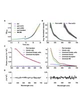
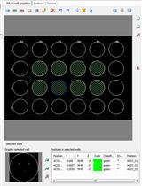
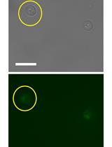
Measuring Mitochondrial ROS in Mammalian Cells with a Genetically Encoded Protein Sensor

用基因编码的蛋白质传感器测定哺乳动物细胞线粒体中的ROS

XZ Xin Zhang
CG Christine Silvia Gibhardt
SC Sabrina Cappello
KZ Katharina Maria Zimmermann
AV Adina Vultur
IB Ivan Bogeski
9967 Views
Jan 20, 2018
Reactive oxygen species (ROS) are not only known for their toxic effects on cells, but they also play an important role as second messengers. As such, they control a variety of cellular functions such as proliferation, metabolism, differentiation and apoptosis. Thus, ROS are involved in the regulation of multiple physiological and pathophysiological processes. It is now apparent that there are transient and local changes in ROS in the cell; in so-called ‘microdomains’ or in specific cellular compartments, which affect signaling events. These ROS hotspots need to be studied in more depth to understand their function and regulation. Therefore, it is necessary to identify and quantify redox signals in single cells with high spatial and temporal resolution. Genetically encoded fluorescence-based protein sensors provide such necessary tools to examine redox-signaling processes. A big advantage of these sensors is the possibility to target them specifically. Mitochondria are essential for energy metabolism and are one of the major sources of ROS in mammalian cells. Therefore, the evaluation of redox potential and ROS production in these organelles is of great interest. Herein, we provide a protocol for the real-time visualization of mitochondrial hydrogen peroxide (H2O2) using the H2O2-specific ratiometric sensor mitoHyPer in adherent mammalian cells.
光漂白后荧光恢复技术分析哺乳动物细胞外泌体转移

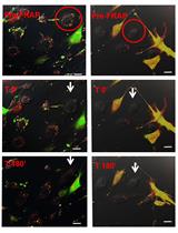
Analysis of Exosome Transfer in Mammalian Cells by Fluorescence Recovery after Photobleaching

光漂白后荧光恢复技术分析哺乳动物细胞外泌体转移

HG Hernán González-King
NG Nahuel A. García
MC María Ciria
Sandra T. Gascón Sandra T. Gascón
RS Rafael S. Sánchez
Helena Grueso Helena Grueso
MG Marta Gómez
RC Rubén M. Cabezuelo
VC Vanesa L. Cava
Pilar Sepúlveda Pilar Sepúlveda
7903 Views
Jan 20, 2018
During the course of evolution, prokaryote and eukaryote cells have developed elegant and to some extent analogous strategies to communicate with each other and to adapt to their surrounding environment. Eukaryotic cells communicate with each other through direct interaction via juxtracrine signaling and/or by secreting soluble factors. These secreted factors can subsequently act on the cell itself (autocrine signaling) or interact with neighboring (paracrine signaling) and distant (endocrine signaling) cells. The transmission of signals between cells and tissues has been traditionally thought to be regulated by a protein-based signaling system. Typically, proteins destined for secretion into the extracellular milieu by exocytosis contain a canonical secretion-targeting sequence (Théry et al., 2002). However, proteins with a non-continuous and stimulus-dependent secretion, proteins that do not contain a canonical secretion-targeting sequence, and species that might be too labile within the extracellular environment (DNA, mRNA, peptides, metabolites, miRNA and other RNA species), can be secreted in small membranous extracellular vesicles (EVs) in a specific manner (Hagiwara et al., 2014). Exosomes represent one broad class of these secreted membrane vesicles with a diameter of 30-130 nm (Cocucci et al., 2009; Théry et al., 2009; Kowal et al., 2014), which are formed inside the secreting cells in endosomal compartments called multivesicular bodies. Molecules loaded into exosomes as well as the intensity of the exosome transfer between cells are important parameters for the subsequent conditioning of recipient cells. Current knowledge on secretion of exosomes and their internalization in recipient cells remains incomplete. It is known that secretion intensity of exosomes varies according to the cellular type and its physiological state (Garcia et al., 2016). Moreover, the different combination of transmembrane proteins on the surface of exosomes that facilitate the adhesion to the cell-extracellular matrix vary the avidity with which a recipient cell captures exosomes (Hoshino et al., 2015). Here, we have developed an in vitro system by which the transfer of exosomes between cells in co-culture can be quantified using FRAP (‘Fluorescence Recovery After Photobleaching’) technology. This protocol has been used to analyze the effects of exosome transfer of hypoxia inducible factor 1-α (HIF-1α) in Mesenchymal Stem Cells (MSC; HIF-MSC) to Human Umbilical Cord Vein Endothelial Cells (HUVEC) (Gonzalez-King et al., 2017).

微生物学

使用CRISPR/Cas9在大肠埃希杆菌中进行逐步多基因敲除

Multiple Stepwise Gene Knockout Using CRISPR/Cas9 in Escherichia coli

使用CRISPR/Cas9在大肠埃希杆菌中进行逐步多基因敲除

EK Enrico König
FZ Francesca Zerbini
IZ Ilaria Zanella
DF Davide Fraccascia
GG Guido Grandi
27508 Views
Jan 20, 2018
With the recent implementation of the CRISPR/Cas9 technology as a standard tool for genome editing, laboratories all over the world are undergoing one of the biggest advancements in molecular biology since PCR. The key advantage of this method is its simplicity and universal applicability for species of any phylum. Of particular interest is the extensively studied Gram-negative bacterium Escherichia coli, as it is considered as the workhorse for both research and industrial purposes. Here, we present a simple, robust and effective protocol using the CRISPR/Cas9 system in combination with the λ Red machinery for gene knockout in E. coli. Crucial in our procedure is the use of a double-stranded donor DNA and a curing strategy for removal of the guide RNA encoding plasmid that allows starting a new mutation after only two working days. Our protocol allows multiple, stepwise gene knockout strains with high mutagenesis efficiencies applicable for high-throughput approaches.
采用Förster共振能量转移法检测大肠埃希杆菌细胞质和周质中蛋白质的相互作用

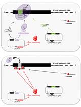
Detection of Protein Interactions in the Cytoplasm and Periplasm of Escherichia coli by Förster Resonance Energy Transfer

采用Förster共振能量转移法检测大肠埃希杆菌细胞质和周质中蛋白质的相互作用

Nils Y. Meiresonne Nils Y. Meiresonne
SA Svetlana Alexeeva
Rv René van der Ploeg
Td Tanneke den Blaauwen
13637 Views
Jan 20, 2018
This protocol was developed to qualitatively and quantitatively detect protein-protein interactions in Escherichia coli by Förster Resonance Energy Transfer (FRET). The described assay allows for the previously impossible in vivo screening of periplasmic protein-protein interactions. In FRET, excitation of a donor fluorescent molecule results in the transfer of energy to an acceptor fluorescent molecule, which will then emit light if the distance between them is within the 1-10 nm range. Fluorescent proteins can be genetically encoded as fusions to proteins of interest and expressed in the cell and therefore FRET protein-protein interaction experiments can be performed in vivo. Donor and acceptor fluorescent protein fusions are constructed for bacterial proteins that are suspected to interact. These fusions are co-expressed in bacterial cells and the fluorescence emission spectra are measured by subsequently exciting the donor and the acceptor channel. A partial overlap between the emission spectrum of the donor and the excitation spectrum of the acceptor is a prerequisite for FRET. Donor excitation can cross-excite the acceptor for a known percentage even in the absence of FRET. By measuring reference spectra for the background, donor-only and acceptor-only samples, expected emission spectra can be calculated. Sensitized emission for the acceptor on top of the expected spectrum can be attributed to FRET and can be quantified by spectral unmixing.
采用LC-MS法鉴定和定量植物相关桔黄假单胞菌和绿针铜绿假单胞菌中次级代谢产物

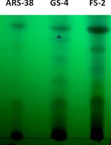
Identification and Quantification of Secondary Metabolites by LC-MS from Plant-associated Pseudomonas aurantiaca and Pseudomonas chlororaphis

采用LC-MS法鉴定和定量植物相关桔黄假单胞菌和绿针铜绿假单胞菌中次级代谢产物

Izzah Shahid Izzah Shahid
MR Muhammad Rizwan
Samina Mehnaz Samina Mehnaz
11643 Views
Jan 20, 2018
Increased antibiotic resistance of plants and human pathogens and continuous use of chemical fertilizers has pushed microbiologists to explore new microbial sources as potential antagonists. In this study, eight strains of Pseudomonas aurantiaca and Pseudomonas chlororaphis, have been isolated from different plant sources and screened for their antagonistic and plant growth promoting potential (Shahid et al., 2017). All strains were compared with reference strain PB-St2 and their secondary metabolites were isolated by the use of solvent partitioning and subjected to LC/ESI/MS for confirmation of compounds. The ESI-mass spectra obtained were used to characterize the surfactants ionization behavior and [M + H]+ and [M + Na]+ ions were monitored for phenazines, derivatives of lahorenoic acid and cyclic lipopeptide (WLIP). LC-MS and HPLC methods were developed to see the elution of dominant metabolites in a single run to avoid the labor and separate methods of detection for all compounds. The method was found suitable and distinctively separated the compounds at different retention times in gradient flow. This method can be helpful to explore the metabolome of Pseudomonas sp. overall and in identification and quantification of strain specific metabolites.
一种分离和纯化噬藻体的液体生物检定方法

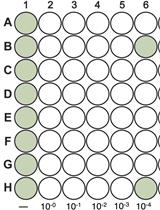
Isolation and Purification of Viruses Infecting Cyanobacteria Using a Liquid Bioassay Approach

一种分离和纯化噬藻体的液体生物检定方法

Caroline Chénard Caroline Chénard
Amy M Chan Amy M Chan
9873 Views
Jan 20, 2018
The following protocol describes the isolation and purification of viruses infecting cyanobacteria using a liquid bioassay approach. Viruses infecting cyanobacteria are also known as cyanophages. This protocol was written specifically for the isolation of cyanophages infecting freshwater cyanobacteria particularly, cyanobacteria that cannot be cultured on solid media. The use of a clonal cyanobacterial culture is recommended for the isolation of viruses. Growth conditions (i.e., media, light cycle and temperature) should be modified based on the host of interest.
细菌 - 真菌对抗和真菌生长预防试验

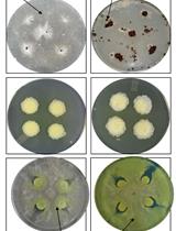
Bacteria-fungal Confrontation and Fungal Growth Prevention Assay

细菌 - 真菌对抗和真菌生长预防试验

Rahul Kumar Rahul Kumar
Durga Madhab Swain Durga Madhab Swain
Sunil Kumar Yadav Sunil Kumar Yadav
IT Isha Tyagi
RK Rajeev Kumar
JD Joyati Das
SG Srayan Ghosh
Gopaljee  Jha Gopaljee Jha
9135 Views
Jan 20, 2018
There are some bacteria which can grow and multiply at the cost of living fungal biomass. They can potentially utilize fungi as a source of nutrients to forage over them. Such phenomenon is known as bacterial mycophagy, however, its mechanistic insights need to be explored to identify the molecules involved in mycophagy for potential utilization in controlling various fungal diseases. Recently we have demonstrated that a rice-associated bacteria Burkholderia gladioli strain NGJ1 exhibits mycophagous ability on several fungi, including Rhizoctonia solani, the necrotrophic fungal pathogen causing sheath blight disease in rice. We hereby describe our validated and efficient methods used to study B. gladioli strain NGJ1-R. solani interactions. These methodologies would be useful for designing assays to study the confrontation between bacteria and fungi which in turn enable discovery of novel antifungal molecules from such bacteria.
哺乳动物正呼肠孤病毒从传染性亚病毒颗粒转化为膜穿透活性颗粒(ISVP-to-ISVP*)分析

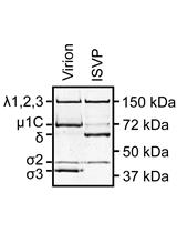
Infectious Subviral Particle to Membrane Penetration Active Particle (ISVP-to-ISVP*) Conversion Assay for Mammalian Orthoreovirus

哺乳动物正呼肠孤病毒从传染性亚病毒颗粒转化为膜穿透活性颗粒(ISVP-to-ISVP*)分析

AS Anthony J. Snyder
PD Pranav Danthi
6030 Views
Jan 20, 2018
The mammalian orthoreovirus (reovirus) outer capsid undergoes a series of conformational changes prior to or during viral entry. These transitions are necessary for delivering the genome-containing core across host cell membranes. This protocol describes an in vitro assay for monitoring the transition into a membrane penetration-active form (i.e., ISVP*).
哺乳动物正呼肠孤病毒传染性亚病毒颗粒诱导的溶血试验

Infectious Subviral Particle-induced Hemolysis Assay for Mammalian Orthoreovirus

哺乳动物正呼肠孤病毒传染性亚病毒颗粒诱导的溶血试验

AS Anthony J. Snyder
PD Pranav Danthi
5647 Views
Jan 20, 2018
Mammalian orthoreovirus (reovirus) utilizes pore forming peptides to penetrate host cell membranes. This step is essential for delivering its genome containing core particle during viral entry. This protocol describes an in vitro assay for measuring reovirus-induced pore formation.
乳酸盐或其他分子诱导的脑膜炎奈瑟氏菌小菌落扩散的活细胞成像

Live-cell Imaging of Neisseria meningitidis Microcolony Dispersal Induced by Lactate or Other Molecules

乳酸盐或其他分子诱导的脑膜炎奈瑟氏菌小菌落扩散的活细胞成像

SS Sara Sigurlásdóttir
OE Olaspers Sara Eriksson
JE Jens Eriksson
AJ Ann-Beth Jonsson
5463 Views
Jan 20, 2018
To efficiently colonize the nasopharyngeal epithelium, the human restricted pathogen Neisseria meningitidis follows a multistep adhesion cascade. First, the bacteria adhere to host cells and aggregate into spherical shaped structures called microcolonies. Several hours later, single bacteria start dispersing from the microcolonies and form a monolayer on top of the host cells. Once in proximity to host cells meningococci can adhere tightly to the epithelial surface or become internalized. This can eventually result in invasion of the mucosal surfaces and gain access to the bloodstream, causing a life-threatening disease. Lactate, a metabolite derived from human epithelial cells, has been previously shown to induce rapid dispersal of N. meningitidis from microcolonies. Here, we describe a host-cell free method based on live-cell imaging to examine the effect of host derived lactate on the timing of N. meningitides microcolony dispersal. Although in this protocol we use lactate, it can be easily modified to test the effects of other molecules.

分子生物学

鉴定酿酒酵母中GFP和mCherry融合蛋白的成熟特征

Characterising Maturation of GFP and mCherry of Genomically Integrated Fusions in Saccharomyces cerevisiae

鉴定酿酒酵母中GFP和mCherry融合蛋白的成熟特征

SS Sviatlana Shashkova
Adam JM Wollman Adam JM Wollman
SH Stefan Hohmann
Mark C  Leake Mark C Leake
8895 Views
Jan 20, 2018
Single-molecule fluorescence microscopy enables unrivaled sub-cellular quantitation of genomically encoded fusions of native proteins with fluorescent protein reporters. Fluorescent proteins must undergo in vivo maturation after expression before they become photoactive. Maturation effects must be quantified during single-molecule analysis. Here we present a method to characterise maturation of GFP and mCherry genetic protein fusions in budding yeast Saccharomyces cerevisiae.

神经科学

急性脑组织切片的制备

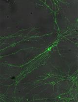
Obtaining Acute Brain Slices

急性脑组织切片的制备

Thomas  Papouin Thomas Papouin
PH Philip G. Haydon
21909 Views
Jan 20, 2018
Obtaining acute brain slices for electrophysiology or amperometric recordings has become a routine procedure in most labs in the field of neuroscience. Yet, protocols describing the step by step process are scarce, in particular for routine acute preparations such as from the mouse hippocampus. Here we provide a detailed protocol for the dissection, extraction and acute slicing of the mouse brain, including tips and list of material required.
成年大鼠颈总动脉闭塞手术作为慢性脑低灌注模型

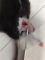
Common Carotid Arteries Occlusion Surgery in Adult Rats as a Model of Chronic Cerebral Hypoperfusion

成年大鼠颈总动脉闭塞手术作为慢性脑低灌注模型

DC Dandan Cao
YB Yunfei Bai
Liang  Li Liang Li
11386 Views
Jan 20, 2018
Chronic cerebral hypoperfusion (CCH) is an important risk factor of vascular dementia (VaD) and Alzheimer’s disease (AD). Hypoxia/ischemia in the whole brain induced by CCH causes serious damage to brain structure and function, which can lead to cognitive impairment. Two-vessel occlusion (2-VO), also known as permanent, bilateral common carotid artery occlusion, is one of the most widely used animal models (e.g., rat) of CCH to investigate the mechanisms of neurodegenerative processes. In this protocol, we present the surgical procedure for 2-VO in rats.
使用FM染料对分离的海马培养神经元中的突触活动进行荧光测定

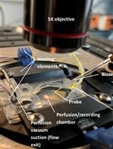
Fluorescent Measurement of Synaptic Activity Using FM Dyes in Dissociated Hippocampal Cultured Neurons

使用FM染料对分离的海马培养神经元中的突触活动进行荧光测定

RL Roman M. Lazarenko
CD Claire E. DelBove
QZ Qi Zhang
10903 Views
Jan 20, 2018
Release and recycling of synaptic vesicles are essential for neurotransmission and synaptic plasticity. To gain mechanistic understanding of these processes, direct measurements of vesicle release and retrieval is indispensable. Styryl dyes like FM1-43 and FM4-64 have been widely used for this purpose and their loading and unloading are reliable measurements for synaptic vesicle release and retrieval in cultured neurons. This protocol describes in detail the procedure of using styryl dyes to label and measure synaptic vesicle uptake and release in cultured rat hippocampal neurons. We also include a brief description of hippocampal culture. In the end, we briefly discuss the commonality and difference among FM dye, pH-sensitive fluorescent proteins and quantum dots in terms of measuring synaptic vesicle behavior.
利用小鼠神经性疼痛模型评估机械性痛觉超敏

Mechanical Allodynia Assessment in a Murine Neuropathic Pain Model

利用小鼠神经性疼痛模型评估机械性痛觉超敏

SN Serena Notartomaso
PS Pamela Scarselli
PD Paola Di Pietro
GB Giuseppe Battaglia
AL Amadeu Llebaria
FC Francisco Ciruela
FN Ferdinando Nicoletti
10811 Views
Jan 20, 2018
Experimental animal models are unique tools (i) to study pain transmission and pathophysiology of neuropathic pain, (ii) to identify novel molecular targets and (iii) to test the potential analgesic effect of specific molecules. The chronic constriction injury (CCI) model of neuropathic pain is the first model of post-traumatic painful peripheral neuropathy, originally developed by Bennett and Xie in the late 1980s. The chronic constriction is performed in the sciatic nerve and induces a partial denervation involving myelinated afferent axons and unmyelinated axons. Damage to unmyelinated axons is much more severe than myelinated afferents. As the model induces a partial denervation, it is very useful for the analysis of pain behaviours. Stimulation of the hind paw, a target of the sciatic nerve, induces pain which can be quantitated. Thus, mechanical allodynia is usually assessed 7, 14 and 21 days after CCI of the sciatic nerve by measuring the hind paw withdrawal response to von Frey filament stimulation. Here, we describe in detail the protocol allowing a reliable and reproducible CCI model in mice. Overall, researchers most commonly use this surgical model to discover more efficacious drugs for the pharmacological control of chronic pain states.
脑切片或其他组织外植体中D-丝氨酸测定

D-serine Measurements in Brain Slices or Other Tissue Explants

脑切片或其他组织外植体中D-丝氨酸测定

Thomas  Papouin Thomas Papouin
PH Philip G. Haydon
7383 Views
Jan 20, 2018
D-serine is an atypical amino acid present in the mammalian body (most amino acids in the mammalian body are L-isomers) that is mostly known in neuroscience for its role as a co-agonist controlling the N-methyl D-aspartate receptor (NMDAR). D-serine levels are decreased in patients with schizophrenia and this is thought to mediate, at least in part, the hypofunction of NMDARs that is central to the glutamate hypothesis for the etiology of this neuropsychiatric disorder. D-serine detection was first established using high performance liquid chromatography, a costly and complex technique that requires high levels of expertise. But with the increasing interest in this unconventional amino acid, there is an increasing need for easier, cheaper and more accessible detection methods. Here we describe the amperometric, biosensor-based method we employed in a recent publication (Papouin et al., 2017b). It allows reliable measurement of D-serine levels from fresh tissue, such as acute brain slices, for concentrations higher than 100 nM, with minimal technical requirements.

植物科学

姜黄素法简单快速测定低浓度硼含量

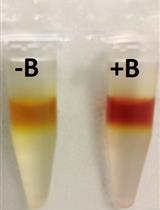
Determination of Boron Content Using a Simple and Rapid Miniaturized Curcumin Assay

姜黄素法简单快速测定低浓度硼含量

Thotegowdanapalya C. Mohan Thotegowdanapalya C. Mohan
AJ Alexandra M. E. Jones
8077 Views
Jan 20, 2018
To determine boron quantity in soil, water and biological samples, several protocols are available. Colorimetric assays are the simplest and cheapest methods which can be used to determine boron concentration. However, published protocols do not give straightforward guidance for beginners to adopt these protocols for routine use in the laboratory. Based on a previously published available procedure, we present a detailed and modified version of a curcumin based colorimetric protocol to determine boron concentration extracted from any sample. Our modified protocol is able to determine up to 0.2 nmole of Boron in a sample volume of 300 µl.

干细胞

小鼠卫星细胞的分离和移植

Mouse Satellite Cell Isolation and Transplantation

小鼠卫星细胞的分离和移植

Elia Angelino Elia Angelino
SR Simone Reano
MF Michele Ferrara
Emanuela Agosti Emanuela Agosti
HS Hana Sustova
Valeria Malacarne Valeria Malacarne
SC Sara Clerici
AG Andrea Graziani
Nicoletta  Filigheddu Nicoletta Filigheddu
10433 Views
Jan 20, 2018
Satellite cell (SC) transplantation represents a powerful strategy to investigate SC biology during muscle regeneration. We described here a protocol for SC isolation from green fluorescent protein (GFP)-expressing mice and their transplantation into murine muscles. This procedure was originally used to assess the effects of the hormone unacylated ghrelin on muscle regeneration, in particular evaluating how the increase of unacylated ghrelin in the recipient muscle affected the engraftment of donor SCs (Reano et al., 2017).
从子宫内膜和经血中分离人子宫内膜干细胞

Human Endometrial Stem Cell Isolation from Endometrium and Menstrual Blood

从子宫内膜和经血中分离人子宫内膜干细胞

Shima Tavakol Shima Tavakol
Freshteh Azedi Freshteh Azedi
Elham Hoveizi Elham Hoveizi
Jafar Ai Jafar Ai
Mohammad Taghi  Joghataei Mohammad Taghi Joghataei
10281 Views
Jan 20, 2018
Human endometrial stem cell/stromal cells (hEnSCs) are isolated from endometrium or menstrual blood and are recognized as a valuable cell type in tissue engineering and cell therapy. Furthermore, hEnSCs, which have CD90 (a mesenchymal marker), CD105 (endoglin), CD44, CD146 (endometrial stem cell markers) and lack CD31 (Endothelial marker), CD34 (hematopoietic marker) and CD133 on the cell surface, are a new source of mesenchymal stem/stromal cells. Additionally, these cells can be encapsulated into self-assembling peptide nanofibers as a 3D scaffold for applications in the treatment of neurodegenerative diseases. Here, we describe a protocol to isolate hEnSCs from endometrium or menstrual blood.