往期刊物2017

卷册: 7, 期号: 18

left

Dec

20

Dec

5

Nov

20

Nov

5

Oct

20

Oct

5

Sep

20

Sep

5

Aug

20

Aug

5

Jul

20

Jul

5

Jun

20

Jun

5

May

20

May

5

Apr

20

Apr

5

Mar

20

Mar

5

Feb

20

Feb

5

Jan

20

Jan

5

right

生物化学

秀丽隐杆线虫胚胎的脂质组学分析

Lipidomic Analysis of Caenorhabditis elegans Embryos

秀丽隐杆线虫胚胎的脂质组学分析

HY Hung-Chi Yang
CH Cheng-Yu Hung
YP Yi-Yun Pan
SL Szecheng J Lo
DC Daniel Tsun-Yee Chiu
9887 Views
Sep 20, 2017
Metabolomic is an emerging field of system biology. Lipidomic, a branch of metabolomic, aims to characterize lipophilic metabolites in biological systems. Caenorhabditis elegans (C. elegans) is a genetically tractable and versatile animal model for novel discovery of lipid metabolism. In addition, C. elegans embryo is simple and homogeneous. Here, we demonstrate detailed procedures of C. elegans culture, embryo isolation, lipid extraction and metabolomic data analysis.
真核先导链/后随链DNA复制的体外分析

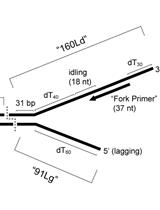
In vitro Assays for Eukaryotic Leading/Lagging Strand DNA Replication

真核先导链/后随链DNA复制的体外分析

GS Grant Schauer
JF Jeff Finkelstein
MO Mike O’Donnell
9312 Views
Sep 20, 2017
The eukaryotic replisome is a multiprotein complex that duplicates DNA. The replisome is sculpted to couple continuous leading strand synthesis with discontinuous lagging strand synthesis, primarily carried out by DNA polymerases ε and δ, respectively, along with helicases, polymerase α-primase, DNA sliding clamps, clamp loaders and many other proteins. We have previously established the mechanisms by which the polymerases ε and δ are targeted to their ‘correct’ strands, as well as quality control mechanisms that evict polymerases when they associate with an ‘incorrect’ strand. Here, we provide a practical guide to differentially assay leading and lagging strand replication in vitro using pure proteins.
苍蝇肠道蛋白酶活性测定

Protease Activity Assay in Fly Intestines

苍蝇肠道蛋白酶活性测定

MN Marie-Paule Nawrot-Esposito
RL Rihab Loudhaief
AG Armel Gallet
8705 Views
Sep 20, 2017
The intestine is a central organ required for the digestion of food, the absorption of nutrients and for fighting against aggressors ingested along with the food. Impairment of gut physiology following mucosal damages impacts its digestive capacities that consequently will affect growth, wellbeing or even survival of the individual. Hence, the assessment of intestinal functions encompasses, among others, the monitoring of its integrity, its cellular renewing, its immune defenses, the production of enteroendocrine hormones and its digestive capacities. Here, we describe in detail how to assess the activity of the proteases secreted in the intestinal lumen of adult Drosophila melanogaster flies. This method can also be used for larval intestines. The present protocol is adapted and improved from the Sigma-Aldrich’s protocol proposed in the ‘Protease Fluorescent Detection Kit’ (Product code PF0100).

癌症生物学

用于监测蒽环类药物进入哺乳动物细胞的摄取分析

Uptake Assays to Monitor Anthracyclines Entry into Mammalian Cells

用于监测蒽环类药物进入哺乳动物细胞的摄取分析

NB Nicolas Brosseau
EA Emil Andreev
Dindial Ramotar Dindial Ramotar
7100 Views
Sep 20, 2017
Anthracyclines, such as doxorubicin and daunorubicin, are DNA damaging agents that autofluoresce and can be readily detected in cells. Herein, we developed suitable assays to quantify and localize daunorubicin in mammalian cells. These assays can be exploited to identify components that are involved in the uptake of anthracyclines.

细胞生物学

电镜冷冻断裂蚀刻技术用于酵母超微结构的简易分析

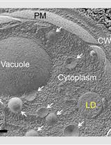
Freeze-fracture-etching Electron Microscopy for Facile Analysis of Yeast Ultrastructure

电镜冷冻断裂蚀刻技术用于酵母超微结构的简易分析

TT Takuma Tsuji
TF Toyoshi Fujimoto
13344 Views
Sep 20, 2017
We describe a streamlined method that enables the quick observation of yeast ultrastructure by electron microscopy (EM). Yeast cells are high-pressure frozen, freeze-fractured to cut across the cytoplasm, and freeze-etched to sublimate ice in the cytosol and the organelle lumen. The cellular structures delineated by these procedures are coated by a thin layer of platinum and carbon deposited by vacuum evaporation, and this platinum–carbon layer, or replica, is observed by transmission EM. The method differs from the deep-etching of pre-extracted samples in that intact live cells are processed without any chemical treatment. Lipid droplets made of unetchable lipid esters are observed most prominently, but other organelles–the nucleus, endoplasmic reticulum, Golgi, vacuoles, mitochondria–and their mutual relationships can be analyzed readily. It is of note that the entire procedure, from quick-freezing to EM observation, can be performed within a day.

免疫学

源自小鼠骨髓的骨髓源性抑制细胞的分化及其与脾树突状细胞的共培养

Differentiation of Myeloid-derived Suppressor Cells from Murine Bone Marrow and Their Co-culture with Splenic Dendritic Cells

源自小鼠骨髓的骨髓源性抑制细胞的分化及其与脾树突状细胞的共培养

GM Giada Mondanelli
CV Claudia Volpi
11443 Views
Sep 20, 2017
Myeloid-derived suppressor cells (MDSCs) possess the ability to suppress the immune response, and to amplify the regulatory properties of other immune cells, i.e., dendritic cells. Here we describe a protocol in which MDSCs were differentiated from murine bone marrow cells, and CD11c+ dendritic cells were purified from murine spleens. MDSCs and CD11c dendritic cells can be co-cultured and the immunoregulatory phenotype of the MDSCs-conditioned dendritic cells could be assessed by means of a specific functional in vivo experiment, i.e., a skin test as a measure of the delayed-type hypersensitivity reaction toward a poorly immunogenic antigen.
一种测定心肌巨噬细胞和心肌成纤维细胞对凋亡细胞吞噬作用的方法

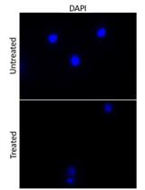
An Assay to Determine Phagocytosis of Apoptotic Cells by Cardiac Macrophages and Cardiac Myofibroblasts

一种测定心肌巨噬细胞和心肌成纤维细胞对凋亡细胞吞噬作用的方法

YH Yuma Horii
SM Shoichi Matsuda
KW Kenji Watari
AN Akiomi Nagasaka
HK Hitoshi Kurose
MN Michio Nakaya
9668 Views
Sep 20, 2017
In myocardial infarction (MI), a number of cardiomyocytes undergo apoptosis. These apoptotic cardiomyocytes are promptly engulfed by phagocytes. If the dead cells are not engulfed, their noxious contents are released outside, resulting in induction of inflammation. Therefore, the removal of these dead cells is necessary. However, the contribution of each phagocyte type to the removal of apoptotic cells in infarcted hearts remains unresolved. Here, we describe an in vitro protocol for a phagocytosis assay to compare the engulfment ability of cardiac macrophages and cardiac myofibroblasts.
心肌成纤维细胞对坏死凋亡细胞的吞噬作用分析

Phagocytosis Assay of Necroptotic Cells by Cardiac Myofibroblasts

心肌成纤维细胞对坏死凋亡细胞的吞噬作用分析

YH Yuma Horii
SM Shoichi Matsuda
KW Kenji Watari
AN Akiomi Nagasaka
HK Hitoshi Kurose
MN Michio Nakaya
7469 Views
Sep 20, 2017
In myocardial infarction (MI), a plenty of cardiomyocytes undergo necrosis and necroptosis due to the lack of oxygen and nutrients. The dead cardiomyocytes are promptly engulfed by phagocytes. When the dead cells are not engulfed, the noxious contents of the cells are released outside, and thus, induce inflammation, and obstruct the function of organs. Therefore, phagocytosis is crucial for maintaining homeostasis of organs. Herein, we describe a protocol of an in vitro phagocytosis assay of necroptotic cells.

微生物学

酿酒酵母中使用人工靶DNA序列进行多重CRISPR/ Cas9的方法

Method for Multiplexing CRISPR/Cas9 in Saccharomyces cerevisiae Using Artificial Target DNA Sequences

酿酒酵母中使用人工靶DNA序列进行多重CRISPR/ Cas9的方法

RG Rachael M. Giersch
Gregory C. Finnigan Gregory C. Finnigan
11883 Views
Sep 20, 2017
Genome manipulation has become more accessible given the advent of the CRISPR (Clustered Regularly Interspaced Short Palindromic Repeats) editing technology. The Cas9 endonuclease binds a single stranded (single guide) RNA (sgRNA) fragment that recruits the complex to a corresponding genomic target sequence where it induces a double stranded break. Eukaryotic repair systems allow for the introduction of exogenous DNA, repair of existing mutations, or deletion of endogenous gene products. Targeting of Cas9 to multiple genomic positions (termed ‘multiplexing’) is achieved by the expression of multiple sgRNAs within the same nucleus. However, an ongoing concern of the CRISPR field has been the accidental targeting of Cas9 to alternative (‘off-target’) DNA locations within a genome. We describe the use (dubbed Multiplexing of Cas9 at Artificial Loci) of installed artificial Cas9 target sequences into the yeast genome that allow for (i) multiplexing with a single sgRNA; (ii) a reduction/elimination in possible off-target effects, and (iii) precise control of the placement of the intended target sequence(s).
果蝇粪便取样

Drosophila Fecal Sampling

果蝇粪便取样

CF Christine Fink
Jv Jakob von Frieling
MK Mirjam Knop
TR Thomas Roeder
9450 Views
Sep 20, 2017
Fecal sampling is a non-invasive method which raises the possibility to study the development and the changes in the microbial community throughout different time points of a fly population or throughout different treatments. This method allows precise manipulation to trigger the fly’s physiology by nutritional interventions, bacterial infections or other stressors. As in most other animals, the intestinal microbiota is essential for a healthy fly-life. Because Drosophila only harbors a relative simple bacterial community with a small variety of round about 8 to 10 different species, it is rather easy to build up the microbial community and to investigate microbial changes after treatment.Another positive effect using the fly’s feces is that bacteria that are not part of the intestinal microbiome, for example Wolbachia, can be excluded directly from the analysis because they are not excreted.Using this method, the generated datasets may reflect a good paradigm to study microbiome associated diseases in a simple fly model or furthermore, to test drugs in a high-throughput approach.

神经科学

在小鼠中缝背核立体定向注射腺相关病毒和埋植套管

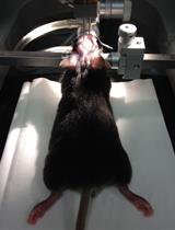
Stereotaxic Adeno-associated Virus Injection and Cannula Implantation in the Dorsal Raphe Nucleus of Mice

在小鼠中缝背核立体定向注射腺相关病毒和埋植套管

PC Patrícia A. Correia
SM Sara Matias
ZM Zachary F. Mainen
18613 Views
Sep 20, 2017
Optogenetic methods are now widespread in neuroscience research. Here we present a detailed surgical procedure to inject adeno-associated viruses and implant optic fiber cannulas in the dorsal raphe nucleus (DRN) of living mice. Combined with transgenic mouse lines, this protocol allows specific targeting of serotonin-producing neurons in the brain. It includes fixing a mouse in a stereotaxic frame, performing a craniotomy, virus injection and fiber implantation. Animals can be later used in behavioral experiments, combined with optogenetic manipulations (Dugué et al., 2014; Correia et al., 2017) or monitoring of neuronal activity (Matias et al., 2017).The described procedure is a fundamental step in both optogenetic and fiber photometry experiments of deep brain areas. It is optimized for serotonin neurons in the DRN, but it can be applied to any other cell type and brain region. When using transgenic mouse lines that express functionally relevant levels of optogenetic tools or reporter lines, the virus injection step can be skipped and the protocol is reduced to the cannula implantation procedure.
胚胎大鼠海马和脑皮质神经元原代培养物的制备

Preparation of Primary Cultures of Embryonic Rat Hippocampal and Cerebrocortical Neurons

胚胎大鼠海马和脑皮质神经元原代培养物的制备

Ivan L. Salazar Ivan L. Salazar
Miranda Mele Miranda Mele
MC Margarida V. Caldeira
 Rui O. Costa Rui O. Costa
BC Bárbara Correia
Simone Frisari Simone Frisari
Carlos B. Duarte Carlos B. Duarte
17592 Views
Sep 20, 2017
This protocol aims at standardizing the procedure to obtain primary cultures of hippocampal and cerebrocortical neurons for in vitro experiments. Cultures should be prepared from cells isolated during embryonic development when neuronal precursor cells are not yet fully differentiated. This helps increasing the quality and quantity of cells, while offering minimal cell death that often occurs during dissociation of differentiated neurons. Cells plated under the appropriate conditions, either in Petri-dishes or in multi-well plates, will develop and establish synaptic contacts over time since the neuronal culture medium provides the nutrients and trophic factors required for differentiation. In this protocol we describe the methodology for the preparation of both cortical and hippocampal neuronal cultures.
自由移动的斑马鱼幼鱼中神经元活性的生物发光监测

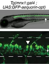
Bioluminescence Monitoring of Neuronal Activity in Freely Moving Zebrafish Larvae

自由移动的斑马鱼幼鱼中神经元活性的生物发光监测

SK Steven Knafo
AP Andrew Prendergast
OT Olivier Thouvenin
SF Sophie Nunes Figueiredo
CW Claire Wyart
9520 Views
Sep 20, 2017
The proof of concept for bioluminescence monitoring of neural activity in zebrafish with the genetically encoded calcium indicator GFP-aequorin has been previously described (Naumann et al., 2010) but challenges remain. First, bioluminescence signals originating from a single muscle fiber can constitute a major pitfall. Second, bioluminescence signals emanating from neurons only are very small. To improve signals while verifying specificity, we provide an optimized 4 steps protocol achieving: 1) selective expression of a zebrafish codon-optimized GFP-aequorin, 2) efficient soaking of larvae in GFP-aequorin substrate coelenterazine, 3) bioluminescence monitoring of neural activity from motor neurons in free-tailed moving animals performing acoustic escapes and 4) verification of the absence of muscle expression using immunohistochemistry.

植物科学

辣椒青霉色素中叶绿素的分离及含量羟化活性的测定

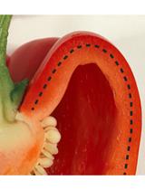
Isolation and Detection of the Chlorophyll Catabolite Hydroxylating Activity from Capsicum annuum Chromoplasts

辣椒青霉色素中叶绿素的分离及含量羟化活性的测定

MH Mareike Hauenstein
SH Stefan Hörtensteiner
7878 Views
Sep 20, 2017
Hydroxylation of chlorophyll catabolites at the so-called C32 position (Hauenstein et al., 2016) is commonly found in all plant species analyzed to date. Here we describe an in vitro hydroxylation assay using Capsicum annuum chromoplast membranes as a source of the hydroxylating activity, which converts the substrate epi-pFCC (epi-primary Fluorescent Chlorophyll Catabolite) (Mühlecker et al., 2000) to epi-pFCC-OH.
在二氨基荧光素(DAF)凝胶上检测植物样品中的蛋白质S-亚硝基硫醇(SNO)

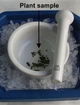
Detection of Protein S-nitrosothiols (SNOs) in Plant Samples on Diaminofluorescein (DAF) Gels

在二氨基荧光素(DAF)凝胶上检测植物样品中的蛋白质S-亚硝基硫醇(SNO)

MR Marta Rodríguez-Ruiz
PM Paulo T. Mioto
José M. Palma José M. Palma
Francisco J. Corpas Francisco J. Corpas
7533 Views
Sep 20, 2017
In plant cells, the analysis of protein S-nitrosothiols (SNOs) under physiological and adverse stress conditions is essential to understand the mechanisms of Nitric oxide (NO)-based signaling. We adapted a previously reported protocol for detecting protein SNOs in animal systems (King et al., 2005) for plant samples. Briefly, proteins from plant samples are separated via non-reducing SDS-PAGE, then the NO bound by S-nitrosylated proteins is released using UV light and, finally, the NO is detected using the fluorescent probe DAF-FM (Rodriguez-Ruiz et al., 2017). Thus, the approach presented here provides a relatively quick and economical procedure that can be used to compare protein SNOs content in plant samples and provide insight in NO-based signaling in plants.