往期刊物2017

卷册: 7, 期号: 16

left

Dec

20

Dec

5

Nov

20

Nov

5

Oct

20

Oct

5

Sep

20

Sep

5

Aug

20

Aug

5

Jul

20

Jul

5

Jun

20

Jun

5

May

20

May

5

Apr

20

Apr

5

Mar

20

Mar

5

Feb

20

Feb

5

Jan

20

Jan

5

right

癌症生物学

葡聚糖硫酸钠(DSS)诱导的结肠炎小鼠模型

Mouse Model of Dextran Sodium Sulfate (DSS)-induced Colitis

葡聚糖硫酸钠(DSS)诱导的结肠炎小鼠模型

Srustidhar Das Srustidhar Das
SB Surinder K. Batra
SR Satyanarayana Rachagani
19092 Views
Aug 20, 2017
Inflammatory bowel disease (IBD) is a chronic inflammatory disease of the intestinal tract and is primarily comprised of Crohn’s disease (CD) and ulcerative colitis (UC). Several murine models that include both chemical induced and genetic models have been developed that mimic some aspects of either CD or UC. These models have been instrumental in our understanding of IBD. Of the chemical induced colitis models, dextran sodium sulfate (DSS) induced colitis model is a relatively simple and very widely used model of experimental colitis.
采用畸胎瘤形成分析法评估多能干细胞的多能性和致瘤性

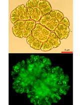
Teratoma Formation Assay for Assessing Pluripotency and Tumorigenicity of Pluripotent Stem Cells

采用畸胎瘤形成分析法评估多能干细胞的多能性和致瘤性

SM Shingo Miyawaki
YO Yohei Okada
HO Hideyuki Okano
KM Kyoko Miura
15378 Views
Aug 20, 2017
Pluripotent stem cells such as induced pluripotent stem cells (iPSCs) and embryonic stem cells (ESCs) form teratomas when transplanted into immunodeficient mice. As teratomas contain all three germ layers (endoderm, mesoderm, ectoderm), teratoma formation assay is widely used as an index of pluripotency (Evans and Kaufman, 1981; Hentze et al., 2009; Gropp et al., 2012). On the other hand, teratoma-forming tumorigenicity also represents a major risk factor impeding potential clinical applications of pluripotent stem cells (Miura et al., 2009; Okano et al., 2013). Recently, we reported that iPSCs derived from naked mole-rat lack teratoma-forming tumorigenicity when engrafted into the testes of non-obese diabetic/severe combined immunodeficient (NOD/SCID) mice due to an ES cell-expressed Ras (ERAS) and Alternative reading frame (ARF)-dependent tumor-suppression mechanism specific to this species (Miyawaki et al., 2016). Here, we describe a method for transplanting pluripotent stem cells into the testes of NOD/SCID mice to generate teratomas for assessing the pluripotency and tumorigenicity.
DNA和有丝分类marker同步染色后利用流式细胞术快速分析细胞周期

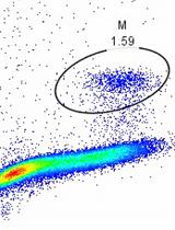
Rapid Profiling Cell Cycle by Flow Cytometry Using Concurrent Staining of DNA and Mitotic Markers

DNA和有丝分类marker同步染色后利用流式细胞术快速分析细胞周期

YS Yuqing Shen
PV Paolo Vignali
RW Ruoning Wang
12998 Views
Aug 20, 2017
The flow cytometric quantitation of DNA content by DNA-binding fluorochrome, propidium iodide (PI) is the most widely used method for cell cycle analysis. However, the commonly used methods are time-consuming and labor-intensive and are incompatible with staining of mitotic markers by fluorescent-labeled antibodies. Here, we report an optimized simple protocol for rapid and simultaneous analysis of characteristic mitotic phosphorylated proteins and DNA content, permitting the quantification of cells in mitosis, G1, S and G2 stage in a single assay. The protocol detailed here employs detergent-based hypotonic solution to rapidly permeabilize cells and allows simultaneous staining of DNA with PI and mitotic marker, phospho-Histone H3, with specific antibody within 20 min. The protocol requires only inexpensive and commercial available reagents and also enables a rapid and complete analysis of cell cycle profile.
乳腺肿瘤类器官中反义寡核苷酸介导的基因敲低技术

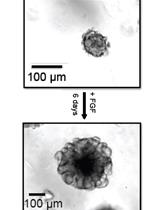
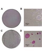
Antisense Oligonucleotide-mediated Knockdown in Mammary Tumor Organoids

乳腺肿瘤类器官中反义寡核苷酸介导的基因敲低技术

Sarah D. Diermeier Sarah D. Diermeier
DS David L. Spector
10306 Views
Aug 20, 2017
Primary mammary tumor organoids grown in 3D are an excellent system to study tumor biology. They resemble the organization and physiology of native epithelia more closely than cancer cell lines grown in 2D, and additionally model interactions with the ECM (Boj et al., 2015; Clevers, 2016; Shamir and Ewald, 2014). Mammary tumor organoids are therefore a promising model system to identify and characterize novel drivers of breast cancer that would be unlikely to be identified using 2D cell lines. Antisense oligonucleotides can be used to efficiently and specifically knockdown target genes in the cell (Bennett et al., 2017). They can be taken up freely by organoids without the need for a transfection agent, making them a convenient tool for routine lab studies and screens.

细胞生物学

小鼠淋巴结基质细胞群的分离和分析

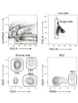
Isolation and Analysis of Stromal Cell Populations from Mouse Lymph Nodes

小鼠淋巴结基质细胞群的分离和分析

YA Yannick O. Alexandre
Scott N Mueller Scott N Mueller
10429 Views
Aug 20, 2017
Our protocol describes a simple procedure for isolating stromal cells from lymph nodes (LN). LN are disrupted then enzymatically digested with collagenase and dispase to produce a single cell suspension that can be stained with fluorescently labelled antibodies and analysed by flow cytometry. This protocol will enable identification of fibroblastic reticular cells (FRC), lymphatic endothelial cells (LEC), blood endothelial cells (BEC) as PNAd+ BEC that form LN high endothelial venules (HEV). This method can be applied to examine LN stromal cell responses during inflammatory events induced by infections or immunologic adjuvants and to subset most leukocytes found in LN.
用于转基因大鼠生产的改良输卵管移植手术

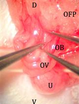
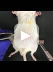
Improved Oviduct Transfer Surgery for Genetically Modified Rat Production

用于转基因大鼠生产的改良输卵管移植手术

LL Laura J. Lambert
LJ Larry W. Johnson
DK Daniel Kennedy
JC Joan Cadillac
Robert A. Kesterson Robert A. Kesterson
9687 Views
Aug 20, 2017
Rat embryo transfer surgeries are becoming more common with targeted nucleases increasing the demand for rat models. This protocol details pre-surgical preparation, improved surgical techniques for placing embryos into the oviduct, and post-surgical care of rats to parturition. Direct application of mouse oviduct transfer protocols results in limited success in the rat. By combining techniques from several widely used protocols in the field, increased yield of live pups born to healthy dams is possible. This protocol is distinct from previously published protocols by the use of a modified anesthesia protocol (Smith et al., 2004), use of analgesia, the addition of peritoneal sutures (Filipiak and Saunders, 2006), incision location and number of transfers per animal (Krinke et al., 2000).

免疫学

.内脏脂肪组织来源基质血管细胞的分离和分析

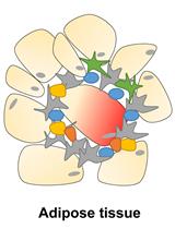
Isolation and Analysis of Stromal Vascular Cells from Visceral Adipose Tissue

.内脏脂肪组织来源基质血管细胞的分离和分析

JV Jessica Vu
WY Wei Ying
13302 Views
Aug 20, 2017
The obesity epidemic is the underlying driver of the type 2 diabetes mellitus epidemic. A remarkable accumulation of various pro-inflammatory immune cells in adipose tissues is a hallmark of obesity and leads to pathogenesis of tissue inflammation and insulin resistance. Here, we describe a detailed protocol to isolate adipose tissue stromal vascular cells (SVCs), which enrich various immune cells of adipose tissues. These SVCs can be used to examine the population and activation status of immune cells by tracking their cell surface antigens, gene expression, and activation of specific signaling pathways.
持续感染仙台病毒的人细胞系的建立

Establishment of a Human Cell Line Persistently Infected with Sendai Virus

持续感染仙台病毒的人细胞系的建立

CC Christopher Coakley
CP Cara Peter
SF Stephanie Fabry
SC Saurabh Chattopadhyay
8413 Views
Aug 20, 2017
Interferon regulatory transcription factor 3 (IRF3) is a transcription factor that upon activation by virus infection promotes the synthesis of antiviral genes, such as the interferons (Hiscott, 2007). In addition to inducing genes, IRF3 triggers antiviral apoptosis by RIG-I-like receptor-induced IRF3 mediated pathway of apoptosis (RIPA), which is independent of its transcriptional activity. RIPA protects against lethal virus infection in cells and mice (Chattopadhyay et al., 2016). In the absence of RIPA, caused by genetic ablation, chemical mutagenesis or inhibition of the pattern recognition receptor (PRR) retinoic acid-inducible gene I (RIG-I), Sendai virus (SeV) infection does not trigger cellular apoptosis and become persistently infected (Peters et al., 2008; Chattopadhyay et al., 2013). IRF3-expressing wild type (WT) cells (U4C) undergo SeV-induced apoptosis; however, the P2.1 cells, which are deficient in IRF3 expression are not capable of triggering viral apoptosis (Figure 1). Ectopic expression of human IRF3 restores the apoptotic activity in P2.1 cells (P2.1/IRF3, Figure 1). SeV is used as a model for studying pathogenic human viruses, which are difficult to work with or require BSL3 facility. We have previously reported that both human and mouse cells can establish SeV persistence in the absence of IRF3’s apoptotic activity (Chattopadhyay et al., 2013). Here, we outline a detailed procedure for the development of a persistently SeV-infected human cell line (Figure 2), which continuously expresses viral protein and produces low levels of infectious viral particles.Figure 1. SeV-induced apoptosis is IRF3-dependent. HT1080-derived cell lines (U4C, P2.1 and P2.1/IRF3) were infected with Sendai virus and three days post infection culture fields were photographed, scale bar represents 50 µm.
使用高内涵显微技术进行巨噬细胞的生存分析

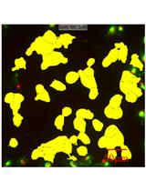
Macrophage Survival Assay Using High Content Microscopy

使用高内涵显微技术进行巨噬细胞的生存分析

RE Remzi Onur Eren
NF Nicolas Fasel
7475 Views
Aug 20, 2017
Macrophages maintain tissue homoeostasis by regulating inflammation and tissue repair mechanisms. Thus, the fate of macrophages has an impact on the state of the tissue. The aim of this protocol is to quantify macrophage survival using high content microscopy and image processing software. Here, we describe a high-content image based protocol to assess the effect of diverse stimuli in combination with pharmacological treatments on macrophage survival in a quantitative, unbiased and high-throughput manner.

微生物学

秀丽隐杆线虫中超氧化物歧化酶(SOD)和过氧化氢酶(CAT)活性测定实验方案

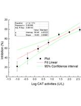
Superoxide Dismutase (SOD) and Catalase (CAT) Activity Assay Protocols for Caenorhabditis elegans

秀丽隐杆线虫中超氧化物歧化酶(SOD)和过氧化氢酶(CAT)活性测定实验方案

JZ Jing Zhang
RC Rui Chen
ZY Zhenyang Yu
LX Lili Xue
37790 Views
Aug 20, 2017
Assays for superoxide dismutase (SOD) and catalase (CAT) activities are widely employed to indicate antioxidant responses underlying the toxic effects of test chemicals. Yet, earlier studies mainly described the procedures as performed according to manufacturer’s instructions without modifications that are specific to any organisms. The present protocol describes the steps in analyzing the superoxide dismutase (SOD) and catalase (CAT) activities in C. elegans, which is a model organism that can be used to study effects of pharmaceutical compounds and environmental pollutants. The main steps include: (1) sample preparation; (2) total protein assay; (3) SOD activity assay; (4) CAT activity assay; and (5) medium list and formula, and also data analysis and performance notes.
利用PEI优化生产,滴定和中和SARS-CoV spike萤光素酶假病毒的方法

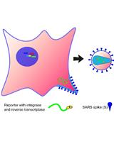
An Optimized Method for the Production Using PEI, Titration and Neutralization of SARS-CoV Spike Luciferase Pseudotypes

利用PEI优化生产,滴定和中和SARS-CoV spike萤光素酶假病毒的方法

GC George Carnell
KG Keith Grehan
FF Francesca Ferrara
EM Eleonora Molesti
NT Nigel Temperton
15774 Views
Aug 20, 2017
The protocol outlined represents a cost-effective, rapid and reliable method for the generation of high-titre viral pseudotype particles with the wild-type SARS-CoV spike protein on a lentiviral vector core using the widely available transfection reagent PEI. This protocol is optimized for transfection in 6-well plates; however it can be readily scaled to different production volumes according to application. This protocol has multiple benefits including the use of readily available reagents, consistent, high pseudotype virus production Relative Luminescence Units (RLU) titres and rapid generation of novel coronavirus pseudotypes for research into strain variation, tropism and immunogenicity/sero-prevalence.
用可产生大肠杆菌毒素的大肠埃希杆菌菌株感染HeLa细胞的实验方案及诱导产生DNA损伤的定量

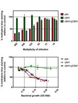
Protocol for HeLa Cells Infection with Escherichia coli Strains Producing Colibactin and Quantification of the Induced DNA-damage

用可产生大肠杆菌毒素的大肠埃希杆菌菌株感染HeLa细胞的实验方案及诱导产生DNA损伤的定量

NB Nadège Bossuet-Greif
MB Marcy Belloy
MB Michèle Boury
EO Eric Oswald
JN Jean-Philippe Nougayrede
11989 Views
Aug 20, 2017
Strains of Escherichia coli bearing the pks genomic island synthesize the genotoxin colibactin. Exposure of eukaryotic cells to E. coli producing colibactin induces DNA damages, ultimately leading to cell cycle arrest, senescence and death. Here we describe a simple method to demonstrate the genotoxicity of bacteria producing colibactin following a short infection of cultured mammalian cells with pks+ E. coli.
使用多重锁式探针检测病原体和氨苄西林耐药基因

Detection of Pathogens and Ampicillin-resistance Genes Using Multiplex Padlock Probes

使用多重锁式探针检测病原体和氨苄西林耐药基因

Rick Conzemius Rick Conzemius
IB Ivan Barišić
9355 Views
Aug 20, 2017
Diagnostic assays for pathogen identification and characterization are limited either by the number of simultaneously detectable targets, which rely on multiplexing methods, or by time constraints due to cultivation-based techniques. We recently presented a 100-plex method for human pathogen characterization to identify 75 bacterial and fungal species as well as 33 clinically relevant β-lactamases (Barišić et al., 2016). By using 16S rRNA gene sequences as barcode elements in the padlock probes, and two different fluorescence channels for species and antibiotic resistance identification, we managed to cut the number of microarray probes needed by half. Consequently, we present here the protocol of an assay with a runtime of approx. 8 h and a detection limit of 105 cfu ml-1. A total of 89% of β-lactamases and 93.7% of species were identified correctly.
利用合理化诱变和X射线晶体学分析在不同功能状态下的信号复合体DesK:DesR

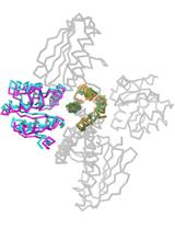
Snapshots of the Signaling Complex DesK:DesR in Different Functional States Using Rational Mutagenesis and X-ray Crystallography

利用合理化诱变和X射线晶体学分析在不同功能状态下的信号复合体DesK:DesR

JI Juan Andres Imelio
NL Nicole Larrieux
AM Ariel Edgardo Mechaly
Felipe Trajtenberg Felipe Trajtenberg
Alejandro Buschiazzo Alejandro Buschiazzo
7435 Views
Aug 20, 2017
We have developed protocols to generate site-specific variants of the histidine-kinase DesK and its cognate response regulator DesR, conducive to trapping different signaling states of the proteins. Co-expression of both partners in E. coli, ensuring an excess of the regulator, was essential for soluble production of the DesK:DesR complexes and further purification. The 3D structures of the complex trapped in the phosphotransferase and in the phosphatase reaction steps, were solved by X-ray crystallography using molecular replacement. The solution was not trivial, and we found that in silico-generated models used as search probes, were instrumental to succeeding in placing a large portion of the complex in the asymmetric unit. Electron density maps were then clear enough to allow for manual model building attaining complete atomic models. These methods contribute to tackling a major challenge in the bacterial signaling field, namely obtaining stable kinase:regulator complexes, in distinct conformational states, amenable for high-resolution crystallographic studies.

分子生物学

使用脉冲追踪法评估蛋白质稳定性的调节

Assessment of Modulation of Protein Stability Using Pulse-chase Method

使用脉冲追踪法评估蛋白质稳定性的调节

ME Mohamed Elgendy
11371 Views
Aug 20, 2017
Pulse-chase technique is a method widely used to assess protein or mRNA stability. The principle of pulse-chase relies on labeling proteins or mRNA produced during a short period of time called ‘pulse’ and then following the rate of disappearance of those labeled proteins over a period of time called ‘chase’. This technique thus allows quantitative analysis of modulation of protein or mRNA stability under different treatments or culturing conditions.

神经科学

使用Hargreaves测试法评估大鼠和小鼠的热疼痛感

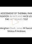
Assessment of Thermal Pain Sensation in Rats and Mice Using the Hargreaves Test

使用Hargreaves测试法评估大鼠和小鼠的热疼痛感

Menghon Cheah Menghon Cheah
JF James W Fawcett
MA Melissa R Andrews
15272 Views
Aug 20, 2017
The Hargreaves test is specifically designed to assess thermal pain sensation in rodents such as rats and mice. This test has been used in experiments involving pain sensitization or recovery of thermal pain response following neural injury and regeneration. We present here a step-by-step protocol highlighted with important notes to guide first-time users through the learning process. Additionally, we have also included representative data from a rat model of sensory denervation showing how the data can be analysed to obtain meaningful results. We hope that this protocol can also assist potential users in deciding whether the Hargreaves test is a suitable test for their experiment.
体外使用寡核苷酸治疗药物对原代皮质神经元mRNA沉默的高通量测定

A High-throughput Assay for mRNA Silencing in Primary Cortical Neurons in vitro with Oligonucleotide Therapeutics

体外使用寡核苷酸治疗药物对原代皮质神经元mRNA沉默的高通量测定

JA Julia F. Alterman
AC Andrew H. Coles
LH Lauren M. Hall
NA Neil Aronin
AK Anastasia Khvorova
MD Marie-Cécile Didiot
10544 Views
Aug 20, 2017
Primary neurons represent an ideal cellular system for the identification of therapeutic oligonucleotides for the treatment of neurodegenerative diseases. However, due to the sensitive nature of primary cells, the transfection of small interfering RNAs (siRNA) using classical methods is laborious and often shows low efficiency. Recent progress in oligonucleotide chemistry has enabled the development of stabilized and hydrophobically modified small interfering RNAs (hsiRNAs). This new class of oligonucleotide therapeutics shows extremely efficient self-delivery properties and supports potent and durable effects in vitro and in vivo. We have developed a high-throughput in vitro assay to identify and test hsiRNAs in primary neuronal cultures. To simply, rapidly, and accurately quantify the mRNA silencing of hundreds of hsiRNAs, we use the QuantiGene 2.0 quantitative gene expression assay. This high-throughput, 96-well plate-based assay can quantify mRNA levels directly from sample lysate. Here, we describe a method to prepare short-term cultures of mouse primary cortical neurons in a 96-well plate format for high-throughput testing of oligonucleotide therapeutics. This method supports the testing of hsiRNA libraries and the identification of potential therapeutics within just two weeks. We detail methodologies of our high throughput assay workflow from primary neuron preparation to data analysis. This method can help identify oligonucleotide therapeutics for treatment of various neurological diseases.
用于定量测定小鼠下丘脑外植体神经肽释放的离体灌流方法

An ex vivo Perifusion Method for Quantitative Determination of Neuropeptide Release from Mouse Hypothalamic Explants

用于定量测定小鼠下丘脑外植体神经肽释放的离体灌流方法

Ophélia Le Thuc Ophélia Le Thuc
Jacques Noël Jacques Noël
Carole Rovère Carole Rovère
7562 Views
Aug 20, 2017
The hypothalamus is a primary brain area which, in mammals, regulates several physiological functions that are all related to maintaining general homeostasis, by linking the central nervous system (CNS) and the periphery. The hypothalamus itself can be considered an endocrine brain region of some sort as it hosts in its different nuclei several kinds of neuropeptide-producing and -secreting neurons. These neuropeptides have specific roles and participate in the regulation of homeostasis in general, which includes the regulation of energy metabolism, feeding behavior, water intake and body core temperature for example. As previously mentioned, in order to exert their effects, these peptides have to be produced but also, and mostly, to be secreted. In this context, it is of great importance to be able to assess how certain conditions, diseases, or treatments can actually influence the secretion of neuropeptides, thus the function of the different neuropeptidergic circuits.One method to assess this is the perifusion of hypothalamic explants followed by quantification of peptides within the collected fractions.Here, we explain step-by-step how to collect fractions during ex vivo perifusion of hypothalamic explants in which one can determine quantitatively neuropeptide/neurohormone release from these viable isolated tissues. Hypothalami perifusion has two great advantages over other existing assays: (1) it allows pharmacological manipulation to dissect out signaling mechanisms underlying release of different neuropeptides/neurohormones in the hypothalamic explants and, (2) it allows simultaneous experiments with different conditions on multiple hypothalami preparations, (3) it is, to our knowledge, the only method that permits the study of neuropeptide secretion in basal conditions and under repeated stimulations with the same hypothalami explants.

植物科学

采用伊文思蓝染色技术定量膜损伤/细胞死亡

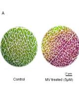
Quantification of Membrane Damage/Cell Death Using Evan’s Blue Staining Technique

采用伊文思蓝染色技术定量膜损伤/细胞死亡

PN Preethi Vijayaraghavareddy
VP Vanitha Adhinarayanreddy
RV Ramu S Vemanna
SM Sheshshayee Sreeman
UM Udayakumar Makarla
19672 Views
Aug 20, 2017
Membrane damage is a hallmark of both biotic and abiotic stress responses. The membrane determines the ability of a cell to sustain altered environmental conditions and hence can be used as a biomarker to assess stress-induced cell damage or death. We present an easy, quick, cost-effective, staining and spectrophotometric method to assess membrane stability of plant cells. In this method, Evan’s blue, an azo dye, is used to assay for cell viability. More specifically, Evan’s blue dye can penetrate through ruptured or destabilized membranes and stain cells. Thus, when plant cells are subjected to stress that compromises membrane integrity, the number of cells that are permeated by Evan’s blue dye will be increased compared to control cells that are not stressed. In contrast, live, healthy cells that are capable of maintaining membrane integrity do not take up Evan’s blue dye. Cells that have taken up Evan’s blue dye will have an accumulation of a blue protoplasmic stain and these stained cells can be qualitatively documented under bright field microscopy with or without the use of a camera. Furthermore, the dye can be extracted from cells that are stained by Evan’s blue dye and can be quantified spectrophotometrically. Using this analysis, the accumulation of dye in positively-stained cells correlates with the extent of cell membrane damage and thus the amount of cells that are stained with Evan’s blue dye under various conditions can be used as an indicator of cellular stress.
各种胁迫条件下根细胞DNA片段化和程序性细胞死亡的TUNEL法评估

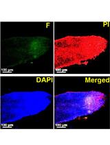
TUNEL Assay to Assess Extent of DNA Fragmentation and Programmed Cell Death in Root Cells under Various Stress Conditions

各种胁迫条件下根细胞DNA片段化和程序性细胞死亡的TUNEL法评估

AT Amit K. Tripathi
Ashwani Pareek Ashwani Pareek
Sneh Lata Singla-Pareek Sneh Lata Singla-Pareek
14260 Views
Aug 20, 2017
DNA damage is one of the common consequences of exposure to various stress conditions. Different methods have been developed to accurately assess DNA damage and fragmentation in cells and tissues exposed to different stress agents. However, owing to the presence of firm cellulosic cell wall and phenolics, plant cells and tissues are not easily amenable to be subjected to these assays. Here, we describe an optimized TUNEL (terminal deoxynucleotidyl transferase-mediated dUTP nick-end labeling) assay-based protocol to determine the extent of DNA fragmentation and programmed cell death in plant root cells subjected to various stress conditions. The method described here has the advantages of simplicity, reliability and reproducibility.
使用硅聚合物印模技术和扫描电子显微镜技术测量气孔的开度、形态和密度

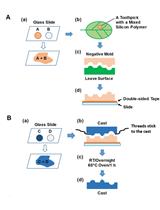
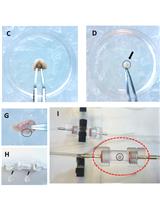
Using Silicon Polymer Impression Technique and Scanning Electron Microscopy to Measure Stomatal Aperture, Morphology, and Density

使用硅聚合物印模技术和扫描电子显微镜技术测量气孔的开度、形态和密度

HW Hui-Chen Wu
YH Ya-Chen Huang
CL Chia-Hung Liu
Tsung-Luo Jinn Tsung-Luo Jinn
9735 Views
Aug 20, 2017
The number of stomata on leaves can be affected by intrinsic development programming and various environmental factors, in addition the control of stomatal apertures is extremely important for the plant stress response. In response to elevated temperatures, transpiration occurs through the stomatal apertures, allowing the leaf to cool through water evaporation. As such, monitoring of stomata behavior to elevated temperatures remains as an important area of research. The protocol allows analysis of stomatal aperture, morphology, and density through a non-destructive imprint of Arabidopsis thaliana leaf surface. Stomatal counts were performed and observed under a scanning electron microscope.
使用CellROX绿色试剂检测布朗葡萄藻集落中的ROS

ROS Detection in Botryococcus braunii Colonies with CellROX Green Reagent

使用CellROX绿色试剂检测布朗葡萄藻集落中的ROS

Edmundo Lozoya-Gloria Edmundo Lozoya-Gloria
IC Ivette Cornejo-Corona
Hem R. Thapa Hem R. Thapa
DB Daniel R. Browne
TD Timothy P. Devarenne
9392 Views
Aug 20, 2017
We analyzed the reactive oxygen species (ROS) accumulation in the colony-forming green microalga Botryococcus braunii in response to several stress inducers such as NaCl, NaHCO3, salicylic acid (SA), methyl jasmonate, and acetic acid. A staining assay using the fluorescent dye CellROX Green was used. CellROX Green is a fluorogenic probe used for measuring oxidative stress in live cells. The dye is weakly fluorescent inside cells in a reduced state but exhibits bright green photostable fluorescence upon oxidation by ROS and subsequent binding to DNA. The large amount of liquid hydrocarbons produced and excreted by B. braunii, creates a highly hydrophobic extracellular environment that makes difficult to study short times defense responses on this microalga. The procedure developed here allowed us to detect ROS in this microalga even within a short period of time (in minutes) after treatment of cells with different stress inducers.

干细胞

从鼠脂肪组织中分离和扩增间充质干细胞

Isolation and Expansion of Mesenchymal Stem Cells from Murine Adipose Tissue

从鼠脂肪组织中分离和扩增间充质干细胞

Natalia V. Andreeva Natalia V. Andreeva
Alexandra A. Dalina Alexandra A. Dalina
Alexander V. Belyavsky Alexander V. Belyavsky
13214 Views
Aug 20, 2017
Mesenchymal stem cells (MSCs) are currently intensively studied due to significant promise which they represent for successful implementations of future cell therapy clinical protocols. This in turn emphasizes importance of careful preclinical studies of MSC effects in various murine disease models. The appropriate cell preparations with reproducible biological properties are important to minimize variability of results of experimental cell therapies. We describe here a simple protocol for isolation of murine MSCs from adipose tissues and their reproducible multi-log expansion under hypoxia conditions.
小鼠胚胎干细胞退出多能性测定实验

Exit from Pluripotency Assay of Mouse Embryonic Stem Cells

小鼠胚胎干细胞退出多能性测定实验

Daniel Cirera-Salinas Daniel Cirera-Salinas
Constance Ciaudo Constance Ciaudo
8903 Views
Aug 20, 2017
A novel method to assess the dissolution of the core pluripotency transcription-factor circuit of mouse Embryonic Stem Cells (mESCs) has been developed (Ying et al., 2003; Betschinger et al., 2013). In order to efficiently identify genes essential for the break-down of the pluripotency network in mutant mESCs with proliferation defects, we adapted this ‘exit from pluripotency assay’ (Bodak et al., 2017; Cirera-Salinas et al., 2017). The protocol described here has been successfully applied to several mESC lines and is easily transposable from one laboratory to another.
人骨髓间充质干细胞共培养实验测定生物力学诱导的TNF-α抑制功效

A Co-culture Assay to Determine Efficacy of TNF-α Suppression by Biomechanically Induced Human Bone Marrow Mesenchymal Stem Cells

人骨髓间充质干细胞共培养实验测定生物力学诱导的TNF-α抑制功效

Miguel F. Diaz Miguel F. Diaz
Siobahn M. Evans Siobahn M. Evans
Scott D. Olson Scott D. Olson
Charles S. Cox Jr Charles S. Cox Jr
Pamela L. Wenzel Pamela L. Wenzel
8778 Views
Aug 20, 2017
The beneficial effects of mesenchymal stem cell (MSC)-based cellular therapies are believed to be mediated primarily by the ability of MSCs to suppress inflammation associated with chronic or acute injury, infection, autoimmunity, and graft-versus-host disease. To specifically address the effects of frictional force caused by blood flow, or wall shear stress (WSS), on human MSC immunomodulatory function, we have utilized microfluidics to model WSS at the luminal wall of arteries. Anti-inflammatory potency of MSCs was subsequently quantified via measurement of TNF-α production by activated murine splenocytes in co-culture assays. The TNF-α suppression assay serves as a reproducible platform for functional assessment of MSC potency and demonstrates predictive value as a surrogate assay for MSC therapeutic efficacy.