往期刊物2017

卷册: 7, 期号: 15

left

Dec

20

Dec

5

Nov

20

Nov

5

Oct

20

Oct

5

Sep

20

Sep

5

Aug

20

Aug

5

Jul

20

Jul

5

Jun

20

Jun

5

May

20

May

5

Apr

20

Apr

5

Mar

20

Mar

5

Feb

20

Feb

5

Jan

20

Jan

5

right

生物化学

秀丽隐杆线虫糖胺聚糖(GAG)的分离和纯化

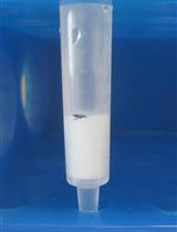
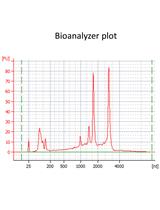
Separation and Purification of Glycosaminoglycans (GAGs) from Caenorhabditis elegans

秀丽隐杆线虫糖胺聚糖(GAG)的分离和纯化

Tabea Dierker Tabea Dierker
LK Lena Kjellén
11714 Views
Aug 5, 2017
The nematode Caenorhabditis elegans is a popular model organism for studies of developmental biology, neurology, ageing and other fields of basic research. Because many developmental processes are regulated by glycosaminoglyans (GAGs) on cell surfaces and in the extracellular matrix, methods to isolate and analyze C. elegans GAGs are needed. Such methods have previously been optimized for other species such as mice and zebrafish. After modifying existing purification protocols, we could recently show that the nematodes also produce chondroitin sulfate, in addition to heparan sulfate, thus challenging the view that only non-sulfated chondroitin was synthesized by C. elegans. We here present our protocol adapted for C. elegans. Since the purification strategy involves separation of non-sulfated and sulfated GAGs, it may also be useful for other applications where this approach could be advantageous.
从哺乳动物细胞中纯化FLAG标记的分泌蛋白

Purification of FLAG-tagged Secreted Proteins from Mammalian Cells

从哺乳动物细胞中纯化FLAG标记的分泌蛋白

EI Eisuke Itakura
CC Changchun Chen
Md Mario de Bono
11138 Views
Aug 5, 2017
This protocol describes a method for purifying glycosylated FLAG-tagged secreted proteins with disulfide bonds from mammalian cells. The purified products can be used for various applications, such as ligand binding assays.
采用膜脂筛查法鉴定生物分子的分子靶点

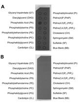
Membrane Lipid Screen to Identify Molecular Targets of Biomolecules

采用膜脂筛查法鉴定生物分子的分子靶点

JJ John R. Jimah
PS Paul H. Schlesinger
NT Niraj H. Tolia
9145 Views
Aug 5, 2017
Proteins that bind to and disrupt cell membranes may target specific phospholipids. Here we describe a protocol to identify the lipid targets of proteins and biomolecules. First, we describe a screen to identify lipids in membranes that are specifically bound by the biomolecule of interest. Second, we describe a method for determining if the presence of these lipids within membranes is necessary for membrane disruption. The methods described here were used to determine that the malaria vaccine candidate CelTOS disrupts cell membranes by specifically targeting phosphatidic acid (Jimah et al., 2016). This protocol has a companion protocol: ‘Liposome disruption assay to examine lytic properties of biomolecules’ which can be applied to examine the ability of the biomolecule to disrupt membranes composed of the lipid target identified by following this protocol (Jimah et al., 2017).
中国蛤蜊中硫酸角质素二糖-支链硫酸软骨素E的分离

Isolation of Keratan Sulfate Disaccharide-branched Chondroitin Sulfate E from Mactra chinensis

中国蛤蜊中硫酸角质素二糖-支链硫酸软骨素E的分离

KH Kyohei Higashi
TT Toshihiko Toida
6921 Views
Aug 5, 2017
Glycosaminoglycans (GAGs) including chondroitin sulfate (CS), dermatan sulfate (DS), heparin (HP), heparan sulfate (HS) and keratan sulfate (KS) are linear, sulfated repeating disaccharide sequences containing hexosamine and uronic acid (or galactose in the case of KS). Recently, a keratan sulfate (KS) disaccharide [GlcNAc6S(β1-3)Galactose(β1-]-branched CS-E was identified from the clam species M. chinensis. Here, we report the isolation protocol for KS-branched CS from M. chinensis.

癌症生物学

RNA干扰筛查鉴定乳腺癌细胞增殖的决定因素

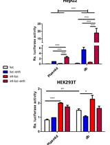
RNA Interference Screening to Identify Proliferation Determinants in Breast Cancer Cells

RNA干扰筛查鉴定乳腺癌细胞增殖的决定因素

YZ Yong-Wei Zhang
RN Rochelle E. Nasto
SJ Sandra A. Jablonski
IS Ilya G Serebriiskii
RS Rishi Surana
JM Joseph Murray
MJ Michael Johnson
RR Rebecca B. Riggins
RC Robert Clarke
EG Erica A. Golemis
LW Louis M. Weiner
9050 Views
Aug 5, 2017
RNAi screening technology has revealed unknown determinants of various biological signaling pathways in biomedical studies. This protocol provided detailed information about how to use RNAi screening to identify proliferation determinants in breast tumor cells. siRNA-based libraries targeting against Estrogen receptor (ER)-network, including 631 genes
relevant to estrogen signaling, was constructed for screening in breast cancer cells. Briefly, reverse transfection of siRNA induced transient gene knockdown in MCF7 cells. First, the transfection reagent for MCF7 cells was selected. Next, the Z’-score assay was used to monitor if screening conditions yielded efficiently. Then, the ER-network siRNA library screening was preceded by automatic machines under optimized experimental conditions.

微生物学

海洋硅藻三角褐指藻中CRISPR / Cas9基因编辑技术

CRISPR/Cas9 Gene Editing in the Marine Diatom Phaeodactylum tricornutum

海洋硅藻三角褐指藻中CRISPR / Cas9基因编辑技术

Marianne Nymark Marianne Nymark
AS Amit Kumar Sharma
MH Marthe C. G. Hafskjold
TS Torfinn Sparstad
AB Atle M. Bones
PW Per Winge
13899 Views
Aug 5, 2017
The establishment of the CRISPR/Cas9 technology in diatoms (Hopes et al., 2016; Nymark et al., 2016) enables a simple, inexpensive and effective way of introducing targeted alterations in the genomic DNA of this highly important group of eukaryotic phytoplankton. Diatoms are of interest as model microorganisms in a variety of areas ranging from oceanography to materials science, in nano- and environmental biotechnology, and are presently being investigated as a source of renewable carbon-neutral fuel and chemicals. Here we present a detailed protocol of how to perform CRISPR/Cas9 gene editing of the marine diatom Phaeodactylum tricornutum, including: 1) insertion of guide RNA target site in the diatom optimized CRISPR/Cas9 vector (pKS diaCas9-sgRNA), 2) biolistic transformation for introduction of the pKS diaCas9-sgRNA plasmid to P. tricornutum cells and 3) a high resolution melting based PCR assay to screen for CRISPR/Cas9 induced mutations.
采用脂质体破碎测定法检测生物分子的裂解性质

Liposome Disruption Assay to Examine Lytic Properties of Biomolecules

采用脂质体破碎测定法检测生物分子的裂解性质

JJ John R. Jimah
PS Paul H. Schlesinger
NT Niraj H. Tolia
13624 Views
Aug 5, 2017
Proteins may have three dimensional structural or amino acid features that suggest a role in targeting and disrupting lipids within cell membranes. It is often necessary to experimentally investigate if these proteins and biomolecules are able to disrupt membranes in order to conclusively characterize the function of these biomolecules. Here, we describe an in vitro assay to evaluate the membrane lytic properties of proteins and biomolecules. Large unilamellar vesicles (liposomes) containing carboxyfluorescein at fluorescence-quenching concentrations are treated with the biomolecule of interest. A resulting increase in fluorescence due to leakage of the dye from liposomes and subsequent dilution in the buffer demonstrates that the biomolecule is sufficient for disrupting liposomes and membranes. Additionally, since liposome disruption may occur via pore-formation or via general solubilization of lipids similar to detergents, we provide a method to distinguish between these two mechanisms. Pore-formation can be identified and evaluated by examining the blockade of carboxyfluorescein release with dextran molecules that fit the pore. The methods described here were used to determine that the malaria vaccine candidate CelTOS and proapoptotic Bax disrupt liposomes by pore formation (Saito et al., 2000; Jimah et al., 2016). Since membrane lipid binding by a biomolecule precedes membrane disruption, we recommend the companion protocol: Jimah et al., 2017.
肽聚糖消化和可溶性片段分析

Digestion of Peptidoglycan and Analysis of Soluble Fragments

肽聚糖消化和可溶性片段分析

Ryan E. Schaub Ryan E. Schaub
Joseph P. Dillard Joseph P. Dillard
12983 Views
Aug 5, 2017
Peptidoglycan (murein) is a vital component of the cell wall of nearly all bacteria, composed of sugars linked by short peptides. This protocol describes the purification of macromolecular peptidoglycan from cultured bacteria and the analysis of enzyme-digested peptidoglycan fragments using high performance liquid chromatography (HPLC). Digested peptidoglycan fragments can be identified by mass spectrometry, or predicted by comparing retention times with other published chromatograms. The quantitative nature of this method allows for the measurement of changes to peptidoglycan composition between different species of bacteria, growth conditions, or mutations. This method can determine the overall architecture of peptidoglycan, such as peptide stem length, the extent of cross-linking, and modifications. Muropeptide analysis has been used to study the function of peptidoglycan-associated proteins and the mechanisms by which bacteria acquire antibiotic resistance.
测定酿酒酵母中淀粉样蛋白诱导氧化应激的白/红色实验方案

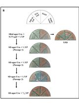
A Protocol of Using White/Red Color Assay to Measure Amyloid-induced Oxidative Stress in Saccharomyces cerevisiae

测定酿酒酵母中淀粉样蛋白诱导氧化应激的白/红色实验方案

VB Vidhya Bharathi
AG Amandeep Girdhar
Basant K Patel Basant K Patel
11375 Views
Aug 5, 2017
The yeast Saccharomyces cerevisiae (S. cerevisiae) harboring ade1 or ade2 mutations manifest red colony color phenotype on rich yeast medium YPD. In these mutants, intermediate metabolites of adenine biosynthesis pathway are accumulated. Accumulated intermediates, in the presence of reduced glutathione, are transported to the vacuoles, whereupon the development of the red color phenotype occurs. Here, we describe a method to score for presence of oxidative stress upon expression of amyloid-like proteins that would convert the red phenotype of ade1/ade2 mutant yeast to white. This assay could be a useful tool for screening for drugs with anti-amyloid aggregation or anti-oxidative stress potency.
利用CRISPR-Cas系统筛选基因组编辑的噬菌体

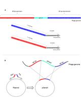
Selection of Genetically Modified Bacteriophages Using the CRISPR-Cas System

利用CRISPR-Cas系统筛选基因组编辑的噬菌体

MM Miriam Manor
Udi Qimron Udi Qimron
11086 Views
Aug 5, 2017
We present a CRISPR-Cas based technique for deleting genes from the T7 bacteriophage genome. A DNA fragment encoding homologous arms to the target gene to be deleted is first cloned into a plasmid. The T7 phage is then propagated in Escherichia coli harboring this plasmid. During this propagation, some phage genomes undergo homologous recombination with the plasmid, thus deleting the targeted gene. To select for these genomes, the CRISPR-Cas system is used to cleave non-edited genomes, enabling isolation of the desired recombinant phages. This protocol allows seamless deletion of desired genes in a T7 phage, and can be expanded to other phages and other types of genetic manipulations as well.
肺炎球菌菌落形态学相位变异的观察

Observation of Pneumococcal Phase Variation in Colony Morphology

肺炎球菌菌落形态学相位变异的观察

Jing Li Jing Li
Juanjuan Wang Juanjuan Wang
FJ Fangfang Jiao
Jing-Ren Zhang Jing-Ren Zhang
9639 Views
Aug 5, 2017
Streptococcus pneumoniae (pneumococcus) is an important human pathogen that causes pneumonia, meningitis, sepsis, and otitis media. This bacterium normally resides in the nasopharynx as a commensal, but sometimes disseminates to sterile sites of humans and causes local or systemic inflammation. This biphasic behavior of S. pneumoniae is correlated with a reversible switch between the opaque and transparent colony forms on agar plates, a phenomenon referred to as phase variation. The opaque variants appear to be more virulent in animal models of bacteremia but are deficient in nasopharyngeal colonization animal models. In contrast, the transparent variants display higher levels of nasopharyngeal colonization but relatively lower virulence in animal models. We have recently demonstrated that pneumococcal phase variation between these two colony types is caused by a reversible switch of genome DNA methylation (or epigenetic) patterns, which is driven by DNA inversions in the DNA methyltransferase genes. Observation of colony morphology is a simple and useful method to differentiate colonies with different characteristics, such as size, color, and opacity. This protocol describes how to study pneumococcal phase variation in colony morphology with a dissection microscope.
极简化哑铃形基因表达载体的优化设计

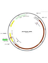
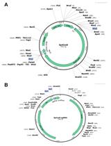
Advanced Design of Minimalistic Dumbbell-shaped Gene Expression Vectors

极简化哑铃形基因表达载体的优化设计

Xiaoou  Jiang Xiaoou Jiang
Volker Patzel Volker Patzel
9552 Views
Aug 5, 2017
Minimal DNA vectors exclusively comprising therapeutically relevant sequences hold great promise for the development of novel therapeutic regimen. Dumbbell-shaped vectors represent non-viral non-integrating DNA minimal vectors which have entered an advanced stage of clinical development (Hardee et al., 2017). Spliceable introns and DNA nuclear import signals such as SV40 enhancer sequences are molecular features that have found multiple applications in plasmid vectors to improve transgene expression. In dumbbells however, effects triggered by introns were not investigated and DNA-based nuclear import sequences have not found applications yet, presumably because dumbbell vectors have continuously been minimized with regard to size. We investigated the effects of an intron and/or SV40 enhancer derived sequences on dumbbell vector driven reporter gene expression. The implementation of a spliceable intron was found to enhance gene expression unconditionally in all investigated cell lines. Conversely, the use of the SV40 enhancer improved gene expression in a cell type-dependent manner. Though both features significantly enlarge dumbbell vector size, neither the intron nor the enhancer or a combination of both revealed a negative effect on gene expression. On the contrary, both features together improved dumbbell-driven gene expression up to 160- or 56-fold compared with plasmids or control dumbbells. Thus, it is highly recommended to consider an intron and the SV40 enhancer for dumbbell vector design. Such an advanced design can facilitate pre-clinical and clinical applications of dumbbell-shaped DNA vectors.
HPLC-MS分析N-乙酰胞壁酸-6-磷酸(MurNAc-6P)的积累

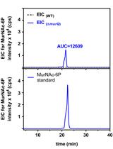
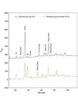
Analysis of N-acetylmuramic acid-6-phosphate (MurNAc-6P) Accumulation by HPLC-MS

HPLC-MS分析N-乙酰胞壁酸-6-磷酸(MurNAc-6P)的积累

MB Marina Borisova
CM Christoph Mayer
8790 Views
Aug 5, 2017
We describe here in detail a high-performance liquid chromatography-mass spectrometry (HPLC-MS)-based method to determine N-acetylmuramic acid-6-phosphate (MurNAc-6P) in bacterial cell extracts. The method can be applied to both Gram-negative and Gram-positive bacteria, and as an example we use Escherichia coli cells in this study. Wild type and mutant cells are grown for a defined time in a medium of choice and harvested by centrifugation. Then the cells are disintegrated and soluble cell extracts are generated. After removal of proteins by precipitation with acetone, the extracts are analyzed by HPLC-MS. Base peak chromatograms of wild type and mutant cell extracts are used to determine a differential ion spectrum that reveals differences in the MurNAc-6P content of the two samples. Determination of peak areas of extracted chromatograms of MurNAc-6P ((M-H)- = 372.070 m/z in negative ion mode) allows quantifying MurNAc-6P levels, that are used to calculate recycling rates of the MurNAc-content of peptidoglycan.
酵母菌中能量依赖性罗丹明6G流出量的测定

Measurement of Energy-dependent Rhodamine 6G Efflux in Yeast Species

酵母菌中能量依赖性罗丹明6G流出量的测定

YG Yvetta Gbelska
NT Nora Toth Hervay
VD Vladimira Dzugasova
AK Alexandra Konecna
8522 Views
Aug 5, 2017
Rhodamine 6G is a highly fluorescent dye often used to determine the transport activity of yeast membrane efflux pumps. The ATP-binding cassette transporter KlPdr5p confers resistance to several unrelated drugs in Kluyveromyces lactis. KlPdr5p also extrudes rhodamine 6G (R6G) from intact yeast cells in an energy-dependent manner. Incubation of yeast cells in the presence of 2-deoxy-D-glucose (inhibitor of glycolysis) and R6G (mitochondrial ATPase inhibitor) leads to marked depletion of intracellular ATP pool (Kolaczkowski et al., 1996). An active KlPdr5p mediated extrusion of R6G from intact yeast cells can be followed by direct measurement of the fluorescence of extruded R6G in the assay buffer.

分子生物学

通过近端dCas9靶向提高CRISPR基因编辑效率

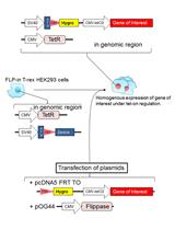
Improving CRISPR Gene Editing Efficiency by Proximal dCas9 Targeting

通过近端dCas9靶向提高CRISPR基因编辑效率

Fuqiang Chen Fuqiang Chen
XD Xiao Ding
YF Yongmei Feng
TS Timothy Seebeck
YJ Yanfang Jiang
GD Gregory D Davis
9991 Views
Aug 5, 2017
Clustered Regularly Interspaced Short Palindromic Repeats (CRISPR) and CRISPR-associated (Cas) systems function as an adaptive immune system in bacteria and archaea for defense against invading viruses and plasmids (Barrangou and Marraffini, 2014). The effector nucleases from some class 2 CRISPR-Cas systems have been repurposed for heterologous targeting in eukaryotic cells (Jinek et al., 2012; Cong et al., 2013; Mali et al., 2013; Zetsche et al., 2015). However, the genomic environments of eukaryotes are distinctively different from that of prokaryotes in which CRISPR-Cas systems have evolved. Mammalian heterochromatin was found to be a barrier to target DNA access by Streptococcus pyogenes Cas9 (SpCas9), and nucleosomes, the basic units of the chromatin, were also found to impede target DNA access and cleavage by SpCas9 in vitro (Knight et al., 2015; Hinz et al., 2015; Horlbeck et al., 2016; Isaac et al., 2016). Moreover, many CRISPR-Cas systems characterized to date often exhibit inactivity in mammalian cells and are thus precluded from gene editing applications even though they are active in bacteria or on purified DNA substrates. Thus, there is a need to devise a means to alleviate chromatin inhibition to increase gene editing efficiency, especially on difficult-to-access genomic sites, and to enable use of otherwise inactive CRISPR-Cas nucleases for gene editing need. Here we describe a proxy-CRISPR protocol for restoring nuclease activity of various class 2 CRISPR-Cas nucleases on otherwise inaccessible genomic sites in human cells via proximal targeting of a catalytically dead Cas9 (Chen et al., 2017). This protocol is exemplified here by using Campylobacter jejuni Cas9 (CjCas9) as nuclease and catalytically dead SpCas9 (SpdCas9) as proximal DNA binding protein to enable CjCas9 to cleave the target for gene editing using single stranded DNA oligo templates.

神经科学

小鼠脑和脊髓中可溶性和不溶性蛋白质组分的提取

Extraction of Soluble and Insoluble Protein Fractions from Mouse Brains and Spinal Cords

小鼠脑和脊髓中可溶性和不溶性蛋白质组分的提取

Oliver Wirths Oliver Wirths
28313 Views
Aug 5, 2017
The current protocol details the preparation of soluble and insoluble protein lysates from mouse brain or spinal cord samples. In detail, tissue homogenization and sequential protein extraction are described. This procedure yields soluble and insoluble protein extracts that can be further processed in down-stream applications like ELISA or Western blotting.
利用小鼠脑和脊髓粗制突触体组分

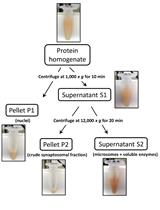
Preparation of Crude Synaptosomal Fractions from Mouse Brains and Spinal Cords

利用小鼠脑和脊髓粗制突触体组分

Oliver Wirths Oliver Wirths
14497 Views
Aug 5, 2017
The current protocol describes the preparation of crude synaptosomal fractions from mouse brain or spinal cord samples. In detail, a sequential protocol yielding crude synaptosomal and light membrane fractions is provided. This fast and easy method might be sufficient to assess the amount of synaptic proteins in down-steam applications like Western-blot or ELISA in e.g., mouse models of Alzheimer’s disease or other neurodegenerative conditions.

植物科学

分离保卫细胞富集组织用于RNA提取

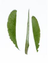
Isolation of Guard-cell Enriched Tissue for RNA Extraction

分离保卫细胞富集组织用于RNA提取

PJ Pirko Jalakas
Dmitry Yarmolinsky Dmitry Yarmolinsky
Hannes Kollist Hannes Kollist
Mikael Brosche Mikael Brosche
15647 Views
Aug 5, 2017
This is a protocol for isolation of guard cell enriched samples from Arabidopsis thaliana plants for RNA extraction. Leaves are blended in ice-water and filtered through nylon mesh to obtain guard cell enriched fragments. With guard cell enriched samples, gene expression analysis can be done, e.g., comparing different gene expression levels in guard cells versus whole leaf to determine if a gene of interest is predominantly expressed in guard cells. It can also be used to study the effect of treatments or different genetic backgrounds in the regulation of the guard cell expressed genes.
拟南芥幼苗中细胞溶质,微粒体,游离多核糖体(FP)和膜结合多聚体(MBPs)的分离

Isolation of Cytosol, Microsome, Free Polysomes (FPs) and Membrane-bound Polysomes (MBPs) from Arabidopsis Seedlings

拟南芥幼苗中细胞溶质,微粒体,游离多核糖体(FP)和膜结合多聚体(MBPs)的分离

Yonghui Zhao Yonghui Zhao
Shengben Li Shengben Li
12047 Views
Aug 5, 2017
The plant endomembrane system plays vital roles for synthesis, modification and secretion of proteins and lipids. From the classic view, only mRNAs encoding secreted proteins could be targeted to the endoplasmic reticulum (ER) for translation via a co-translational translocation manner, however, recently this model has been challenged by accumulative evidence that lots of cytosolic mRNAs could also associate with ER, and that some categories of small RNAs are enriched on ER. These results suggested unrevealed functions of ER beyond our current knowledge. The large scale identification of RNAs and proteins on microsome is crucial to demonstrating the ER function and the studies will be boosted by next generation sequencing technology. This protocol provides a technical workflow to isolate the cytosol, microsome, free polysome (FP) and membrane bound polysome (MBP) from plant tissue. The isolated fractions are suitable for genome wide profiling of mRNAs, small RNAs and proteins.
用于大规模表型分析的小麦胚芽鞘禾谷镰孢菌接种

Wheat Coleoptile Inoculation by Fusarium graminearum for Large-scale Phenotypic Analysis

用于大规模表型分析的小麦胚芽鞘禾谷镰孢菌接种

LJ Lei-Jie Jia
WW Wan-Qiu Wang
Wei-Hua Tang Wei-Hua Tang
10968 Views
Aug 5, 2017
The ascomycete fungus Fusarium graminearum is a destructive fungal pathogen of wheat, barley and maize. Although reverse genetics and homologous recombination gene deletion methods have generated thousands of gene deletion mutants of F. graminearum, evaluating virulence of these fungal mutants is still a rate-limiting step. Here we present a protocol for inoculation of wheat coleoptiles with conidial suspensions for large-scale phenotypic analysis, and describe how it can also be used to assess fungal infectious growth and symptom developmentat a cellular scale. The inoculation method described in this protocol provides highly reproducible results in wheat coleoptile infection by F. graminearum.
腐生与植物内的黑粉病真菌-麦雀生黑粉菌在生长期间高差异表达基因分析

Overrepresentation Analyses of Differentially Expressed Genes in the Smut Fungus Ustilago bromivora during Saprophytic and in planta Growth

腐生与植物内的黑粉病真菌-麦雀生黑粉菌在生长期间高差异表达基因分析

AC Angelika Czedik-Eysenberg
FR Franziska Rabe
HE Heinz Ekker
CC Carmen Czepe
AD Armin Djamei
8892 Views
Aug 5, 2017
We have established the Ustilago bromivora–Brachypodium spp. interaction as a new model pathosystem for biotrophic fungal plant infections of the head smut type (Rabe et al., 2016). In this protocol, the methodology used for comparing gene expression between saprophytic and in planta growth of the fungus is described. The experimental and analytical pipeline, how next generation RNA sequencing (Illumina RNA-Seq) analysis can be used to obtain lists of genes significantly up or down regulated in planta in comparison to axenic culture is given. Furthermore, different methods to identify functional categories that are over- or under-represented among specific classes of genes are presented.
拟南芥幼苗和愈伤组织中多胺和百草枯转运测定

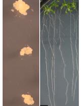
Polyamine and Paraquat Transport Assays in Arabidopsis Seedling and Callus

拟南芥幼苗和愈伤组织中多胺和百草枯转运测定

HC Haoxi Chai
YS Yun Shen
Huazhong Shi Huazhong Shi
7134 Views
Aug 5, 2017
Polyamines (PAs) are polycationic compounds found in all living organisms and play crucial roles in growth and survival. We here show the ‘Polyamine and paraquat (PQ) transport assay’ protocol, which can be used to examine the uptake activity of PA/PQ transporters. We have used this protocol to demonstrate that PUT3 in Arabidopsis is a polyamine transporter and is able to take up spermidine and its analog paraquat.

干细胞

诱导的人多能干细胞(iPS细胞)和胚胎干细胞(ES细胞)向树突状细胞(DC)亚型的分化

Differentiation of Human Induced Pluripotent Stem Cells (iPS Cells) and Embryonic Stem Cells (ES Cells) into Dendritic Cell (DC) Subsets

诱导的人多能干细胞(iPS细胞)和胚胎干细胞(ES细胞)向树突状细胞(DC)亚型的分化

SS Stephanie Sontag
MF Malrun Förster
KS Kristin Seré
Martin Zenke Martin Zenke
15513 Views
Aug 5, 2017
Induced pluripotent stem cells (iPS cells) are engineered stem cells, which exhibit properties very similar to embryonic stem cells (ES cells; Takahashi and Yamanaka, 2016). Both iPS cells and ES cells have an extraordinary self-renewal capacity and can differentiate into all cell types of our body, including hematopoietic stem/progenitor cells and dendritic cells (DC) derived thereof. This makes iPS cells particularly well suited for studying molecular mechanisms of diseases, drug discovery and regenerative therapy (Grskovic et al., 2011; Bellin et al., 2012; Robinton and Daley, 2012).DC are the major antigen presenting cells of the immune system and thus they are key players in modulating and directing immune responses (Merad et al., 2013). DC patrol peripheral and interface tissues (e.g., lung, intestine and skin) to detect invading pathogens, and upon activation they migrate to lymph nodes to activate and prime lymphocytes. DC comprise a phenotypically heterogeneous family with functionally specialized subsets (Schlitzer and Ginhoux, 2014). Generally, classical DC (cDC) and plasmacytoid DC (pDC) are distinguished, exhibiting a classical and plasma cell-like DC morphology, respectively. cDC recognize a multitude of pathogens and secrete proinflammatory cytokines upon activation, while pDC are specialized to detect intracellular pathogens and secrete type I interferons (Merad et al., 2013; Schlitzer and Ginhoux, 2014). cDC are further divided into cross-presenting cDC1 and conventional cDC2, in the human system referred to as CD141+ Clec9a+ cDC1 and CD1c+ CD14- cDC2. Human pDC are characterized as CD303+ CD304+ (Jongbloed et al., 2010; Joffre et al., 2012; Swiecki and Colonna, 2015). To investigate subset specification and function of human DC, we established a protocol to generate cDC1, cDC2 and pDC in vitro from human iPS cells (or ES cells) (Sontag et al., 2017). Therefore, we differentiated iPS cells (or ES cells), via mesoderm commitment and hemato-endothelial specification, into CD43+ CD31+ hematopoietic progenitors. Subsequently, those were seeded onto inactivated OP9 stromal cells with FLT3L, SCF, GM-CSF and IL-4 or FLT3L, SCF and GM-CSF to specify cDC1 and cDC2, or cDC1 and pDC, respectively.
小鼠Müller细胞分离与培养

Mouse Müller Cell Isolation and Culture

小鼠Müller细胞分离与培养

XL Xiao Liu
LT Luosheng Tang
YL Yongqing Liu
12057 Views
Aug 5, 2017
Müller cells are the major supportive and protective glial cells across the retina. Unlike in fish, they have lost the capacity to regenerate the retina in mammals. But, mammalian Müller cells still retain certain retinal stem cell properties with various degree of self-renewal and differentiation potentials, and thereby held a merit in cell-based therapies for treating retinal degeneration diseases. In our laboratory, we use an enzymatic procedure to isolate, purify, and culture mouse Müller cells.
秀丽隐杆线虫肿瘤生殖细胞系突变体生殖细胞的原代培养系统

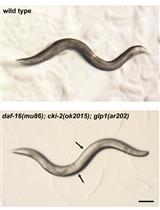
Primary Culture System for Germ Cells from Caenorhabditis elegans Tumorous Germline Mutants

秀丽隐杆线虫肿瘤生殖细胞系突变体生殖细胞的原代培养系统

AV Alexandra S. Vagasi
MR Mohammad M. Rahman
SC Snehal N. Chaudhari
EK Edward T. Kipreos
8662 Views
Aug 5, 2017
The Caenorhabditis elegans germ line is an important model system for the study of germ stem cells. Wild-type C. elegans germ cells are syncytial and therefore cannot be isolated in in vitro cultures. In contrast, the germ cells from tumorous mutants can be fully cellularized and isolated intact from the mutant animals. Here we describe a detailed protocol for the isolation of germ cells from tumorous mutants that allows the germ cells to be maintained for extended periods in an in vitro primary culture. This protocol has been adapted from Chaudhari et al., 2016.