往期刊物2015

卷册: 5, 期号: 5

left

Dec

20

Dec

5

Nov

20

Nov

5

Oct

20

Oct

5

Sep

20

Sep

5

Aug

20

Aug

5

Jul

20

Jul

5

Jun

20

Jun

5

May

20

May

5

Apr

20

Apr

5

Mar

20

Mar

5

Feb

20

Feb

5

Jan

20

Jan

5

right

免疫学

采用流式细胞术分离脾脏中树突细胞

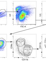
Isolation of Splenic Dendritic Cells Using Fluorescence-activated Cell Sorting

采用流式细胞术分离脾脏中树突细胞

ST Simon J Tavernier
FO Fabiola Osorio
SJ Sophie Janssens
Bart N Lambrecht Bart N Lambrecht
17289 Views
Mar 5, 2015
The spleen is a vastly vasculated organ and consists of a complex organized network of innate and adaptive immune cells. This permits the specialized functions of the spleen such as antibacterial and antifungal immunity and iron metabolism among others (Mebius and Kraal, 2005). Different dendritic cell (DC) subsets reside in the spleen and can be defined by the expression of unique surface markers. These DC subsets are recognized to perform non-redundant functions in the immune system (Merad et al., 2013). In our recent study, we found that Inositol Requiring Enzyme (IRE)-1 is specifically activated in splenic CD8a+ DCs. Furthermore, loss of X-box binding protein (XBP)-1 – the transcription factor regulated by IRE-1 – resulted in defective cross-presentation of dead cell associated antigens by splenic CD8a+ DCs (Osorio et al., 2014). This protocol allows the isolation of specific DC subsets for experimental use ex-vivo.

微生物学

枯草杆菌生物膜的囊泡分离

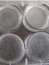
Vesicle Isolation from Bacillus subtilis Biofilm

枯草杆菌生物膜的囊泡分离

Lisa Brown Lisa Brown
AK Anne Kessler
AC Arturo Casadevall
12911 Views
Mar 5, 2015
Bacterial biofilms are associated clinically with many bacterial infections including those caused by bacteria such as Pseudomonas aeruginosa and Staphylococcus aureus. In recent years, extracellular vesicles produced by bacteria have been isolated from biofilm communities. Vesicles have been described in depth and can encapsulate various virulence factors including toxins and immunomodulatory compounds. Vesicles may be important for virulence and survival by serving as a vehicle for the secretion and concentrated delivery of these molecules. Studying extracellular vesicles is an important step towards understanding biofilm formation, structure, and disruption with the ultimate goal of preventing or treating hospital infections caused by bacterial pathogens residing in biofilms. Here we describe the protocol for isolating vesicles from biofilm produced by Bacillus subtilis.
采用Glucuronoxylomannan免疫胶体金透射电子显微镜法检测枯草杆菌中脂质聚合自然产生的胞外膜囊泡的分化

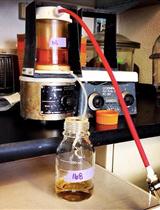
Differentiation of Naturally Produced Extracellular Membrane Vesicles from Lipid Aggregation by Glucuronoxylomannan Immunogold Transmission Electron Microscopy in Bacillus subtilis

采用Glucuronoxylomannan免疫胶体金透射电子显微镜法检测枯草杆菌中脂质聚合自然产生的胞外膜囊泡的分化

Lisa Brown Lisa Brown
GP Geoff Perumal
AC Arturo Casadevall
10513 Views
Mar 5, 2015
Recently, membrane vesicle (MV) production was described in Gram-positive bacteria, which harbor a variety of components such as toxins, antibiotic resistance proteins, proteases, DNA, and immune modulators. Free lipids have the ability to form micelles, thus it is important to rule out spontaneous association of lipids into vesicle-like structures and rather, that MVs are produced naturally by a metabolically active cell. Here, we describe a protocol utilizing the polysaccharide, glucuronoxylomannan (GXM) from Cryptococcus neoformans (C. neoformans) as a marker to differentiate naturally produced MVs from vesicles that form spontaneously in the Gram-positive model organism, Bacillus subtilis (B. subtilis). MVs are purified from bacterial cultures grown in the presence of GXM; MVs naturally produced by cells would not contain GXM in the lumen whereas vesicular structures forming in the media could encapsulate GXM and this can be visualized via immunogold transmission electron microscopy.
气相色谱质谱双极分析法体外测定肌醇3磷酸盐合成酶(INO1)的活性

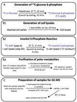
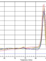
A Gas Chromatography-Mass Spectrometry-Based Two Stage Assay for Measurement of in vitro myo-Inositol 3-phosphate Synthase (INO1) Activity

气相色谱质谱双极分析法体外测定肌醇3磷酸盐合成酶(INO1)的活性

JM James I. MacRae
Malcolm J. McConville Malcolm J. McConville
9615 Views
Mar 5, 2015
This method describes an in vitro assay for measuring INO1 enzyme activity (the conversion of glucose 6-phosphate to myo-inositol 3-phosphate) in cell-free extracts. The method was first described for Plasmodium falciparum cells in MacRae et al. (2014) and consists of two parts: Part 1 describes the assay itself while part 2 describes analysis of the myo-inositol 3-phosphate product using gas chromatography-mass spectrometry (GC-MS).

神经科学

成年斑马鱼30秒应激实验

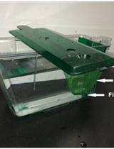
Thirty-Second Net Stressor Task in Adult Zebrafish

成年斑马鱼30秒应激实验

Steven Tran Steven Tran
Robert Gerlai Robert Gerlai
8986 Views
Mar 5, 2015
Zebrafish have become a popular animal model for behavioral neuroscience (Gerlai, 2014). Recent studies have demonstrated that brief experimental handling prior to euthanizing animals can subsequently alter biological measures quantified post-mortem (e.g. cortisol levels) (Ramsay et al., 2009; Tran et al., 2014). Here we provide a detailed protocol for a simple 30-sec net stressor task for adult zebrafish that increases whole-body cortisol levels without altering the levels of whole-brain dopamine, 3, 4-dihydroxyphenylacetic acid, serotonin, and 5-hydroxyindoleacetic acid (Tran et al., 2014).

植物科学

水稻叶肉细胞原生质体分离和细胞核分级分离的有效方法

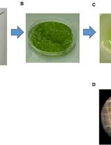
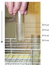
An Efficient Procedure for Protoplast Isolation from Mesophyll Cells and Nuclear Fractionation in Rice

水稻叶肉细胞原生质体分离和细胞核分级分离的有效方法

Mehdi Jabnoune Mehdi Jabnoune
DS David Secco
CL Cécile Lecampion
CR Christophe Robaglia
QS Qingyao Shu
YP Yves Poirier
24245 Views
Mar 5, 2015
Plant protoplasts, a proven physiological and versatile cell system, are widely used in high-throughput analysis and functional characterization of genes. Green protoplasts have been successfully used in investigations of plant signal transduction pathways related to hormones, metabolites and environmental challenges. This protocol, adapted from Zhang et al. (2011), describes a procedure for the isolation of rice protoplasts from green tissue and shows an efficient and rapid method for isolation of nuclei form these protoplasts which are commonly used in a variety of experimental procedures including the isolation of high-molecular-weight DNA (Watson and Thompson, 1986), in vitro DNA synthesis (Roman, 1980), isolation of labeled transcripts for differential screening of cDNA libraries (Somssich et al., 1989), preparation of nuclear extracts for in vitro transcription systems (Roberts and Okita, 1991), isolation of nuclear proteins (Harrison et al., 1992) and studies of protein targeting to the nucleus (Hicks and Raikhel, 1993).
柑橘类水果中的抗坏血酸提取及高效液相色谱法测定

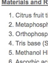
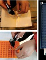
Citrus Fruit Ascorbic Acid Extraction and Quantification by HPLC

柑橘类水果中的抗坏血酸提取及高效液相色谱法测定

Enriqueta Alós Enriqueta Alós
Joanna Lado Joanna Lado
María Jesús Rodrigo María Jesús Rodrigo
Lorenzo Zacarías Lorenzo Zacarías
19007 Views
Mar 5, 2015
Citrus are among the most relevant sources of vitamin C (ascorbic acid + dehydroascorbic acid). Recent studies have revealed that it increases in the peel as fruit ripens and remains constant or even decreases in the pulp tissue. Moreover, important differences on ascorbic acid content exist among citrus varieties in both tissues. Here we describe a simple method for vitamin C analysis/quantification in the peel and pulp tissues of citrus fruit.
分离水稻固体组织的多核糖体结合mRNA用于RT-PCR和图谱实验

Isolation of Polysome-bound mRNA from Rice Solid Tissues Amenable for RT-PCR and Profiling Experiments

分离水稻固体组织的多核糖体结合mRNA用于RT-PCR和图谱实验

Mehdi Jabnoune Mehdi Jabnoune
DS David Secco
CL Cécile Lecampion
CR Christophe Robaglia
QS Qingyao Shu
YP Yves Poirier
14981 Views
Mar 5, 2015
Polysome profile analysis is a frequently performed task in translational control research that not only enables direct monitoring of the efficiency of translation but can easily be extended with a wide range of downstream applications such as Northern and western blotting, genome-wide microarray analysis or qRT-PCR. Here, we describe a method for the isolation and quantification of high-quality polysome-bound mRNA complexes from small quantities of liquid-nitrogen-frozen solid tissue samples of rice shoots/roots. The mRNA obtained can be further analyzed by methods that evaluate polysomal mRNA abundance at the individual transcript or global level.
黑吉豆中小RNA的提取和miRNA的定量PCR

Extraction of Small RNA and qPCR Validation of miRNAs in Vigna mungo

黑吉豆中小RNA的提取和miRNA的定量PCR

Sujay Paul Sujay Paul
Anirban Kundu Anirban Kundu
Amita Pal Amita Pal
10949 Views
Mar 5, 2015
Small RNAs like microRNAs (miRNAs), small interfering RNAs (siRNAs) and other noncoding RNAs including snRNA and snoRNA have tremendous impact on eukaryotic gene regulation. Extraction of high quality small RNAs is an important prerequisite for experimental analyses of miRNAs. This will prevent RNA degradation and remove associated contaminations including polyphenols, polysaccharides and other secondary metabolites. In this protocol we describe a simple way to isolate small RNAs from the leaf tissues of Vigna mungo combining the protocols of two commercially available kits with some modifications.
采用活体细胞成像法观察氧化还原敏感性GFP(roGFP)测量花粉中的氧化还原反应

Measurement of Cellular Redox in Pollen with Redox-Sensitive GFP (roGFP) Using Live Cell Imaging

采用活体细胞成像法观察氧化还原敏感性GFP(roGFP)测量花粉中的氧化还原反应

Wei-Jie Huang Wei-Jie Huang
Wei-Hua Tang Wei-Hua Tang
10351 Views
Mar 5, 2015
Redox homeostasis is a fundamental property of living cells and responds actively to both cellular metabolism and external stimulus. The development of redox-sensitive GFP (roGFP) enables dynamic monitoring of changes in cellular redox poise (Hanson et al., 2014). When excited alternatively at 405 nm and 488 nm, these probes exhibit significant opposing shifts at the emission spectra (505-530 nm), which enables ratiometric measurement of relative redox values. A more oxidized environment results in a higher 405/488 ratio. Previously, successful application of roGFPs in leaf epidermis or root cells has been reported. Here we provide a protocol describing the application of roGFP1 imaging in growing pollen tubes by confocal laser scanning microscopy.
直接测序法检测RNA 编辑

RNA Editing Detection by Direct Sequencing

直接测序法检测RNA 编辑

Mehdi Jabnoune Mehdi Jabnoune
DS David Secco
CL Cécile Lecampion
CR Christophe Robaglia
QS Qingyao Shu
YP Yves Poirier
9020 Views
Mar 5, 2015
RNA editing is a widespread post-transcriptional phenomenon through which primary RNA sequences are altered by nucleotide insertion/deletion or base conversion. It occurs in a variety of organisms and cooperates with alternative splicing in increasing both proteomic and transcriptomic complexity. We describe here a method allowing RNA editing events detection by performing direct sequencing of both genomic DNA and cDNA from the same source.