往期刊物2015

卷册: 5, 期号: 1

left

Dec

20

Dec

5

Nov

20

Nov

5

Oct

20

Oct

5

Sep

20

Sep

5

Aug

20

Aug

5

Jul

20

Jul

5

Jun

20

Jun

5

May

20

May

5

Apr

20

Apr

5

Mar

20

Mar

5

Feb

20

Feb

5

Jan

20

Jan

5

right

生物化学

小鼠排泄物中的脂质提取

Lipid Extraction from Mouse Feces

小鼠排泄物中的脂质提取

Daniel Kraus Daniel Kraus
QY Qin Yang
BK Barbara B. Kahn
17965 Views
Jan 5, 2015
The analysis of feces composition is important for the study of energy metabolism, which comprises various measurements of energy intake, energy expenditure, and energy wasting. The current protocol describes how to measure energy-dense lipids in mouse feces using a modification of the method proposed by Folch et al. (1957).

癌症生物学

放线菌酮追踪检测分析蛋白质的稳定性

Analysis of Protein Stability by the Cycloheximide Chase Assay

放线菌酮追踪检测分析蛋白质的稳定性

SK Shih-Han Kao
WW Wen-Lung Wang
CC Chi-Yuan Chen
YC Yih-Leong Chang
YW Yi-Ying Wu
YW Yi-Ting Wang
SW Shu-Ping Wang
AN Alexey I Nesvizhskii
YC Yu-Ju Chen
TH Tse-Ming Hong
Pan-Chyr Yang Pan-Chyr Yang
62497 Views
Jan 5, 2015
Comparison of protein stability in eukaryotic cells has been achieved by cycloheximide, which is an inhibitor of protein biosynthesis due to its prevention in translational elongation. It is broadly used in cell biology in terms of determining the half-life of a given protein and has gained much popularity in cancer research. Here we present a full cycloheximide chase assay in our laboratory using a lung adenocarcinoma cell line, CL1-5, as a model.
三维球状体共培养侵袭研究

Three Dimensional Spheroid Co-culture Invasion Assay

三维球状体共培养侵袭研究

EV Eldo T Verghese
TH Thomas A Hughes
13191 Views
Jan 5, 2015
The assay was developed to investigate the impact of stromal cells of different types (in our case breast cancer associated fibroblasts stably manipulated to modify expression of genes of interest) on the invasive capacity of epithelial cancer cells (in our case breast cancer cell lines) (Verghese et al., 2013). Typical two dimensional invasion assays do necessarily account for the presence of extracellular matrix that is present around the stromal and tumour cells in vivo and therefore cellular behaviour within these cultures may be non-physiological. This spheroid assay was developed to attempt to replicate more closely the environment that is present around breast cancer stromal and tumour cells in actual tumours (Verghese et al., 2013). Extra cellular matrix composed of both collagen IV and collagen I is included and fibroblasts and epithelial cells are given the opportunity to develop “physiological” interactions (Verghese et al., 2013; Hooper et al., 2006). The method was developed from Nowicki et al. (2008), and we have published data using it in Verghese et al. (2013).

免疫学

适用于CyTOF质谱流式细胞仪的外周血单核细胞(PBMC)细胞因子胞内染色方法

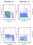
Intracellular Cytokine Staining on PBMCs Using CyTOFTM Mass Cytometry

适用于CyTOF质谱流式细胞仪的外周血单核细胞(PBMC)细胞因子胞内染色方法

DL Dongxia Lin
SG Sheena Gupta
Holden Maecker Holden Maecker
23499 Views
Jan 5, 2015
In this protocol, we use a CyTOFTM mass cytometry to collect single-cell data on a large number of cytokines/chemokines as well as cell-surface proteins that characterize T cells and other immune cells. The current selected mass window in AW 103-203 includes the lanthanides used for most antibody labeling, along with iridium and rhodium for DNA intercalators. The output data are in the format as .txt and .fcs files, which is compatible with many analysis programs. This protocol could be adapted to include tetramers into the staining panel, but we have not optimized for that purpose. The principal steps of intracellular cytokine staining are as follows: First, cells are activated for a few hours using either a specific peptide or a non-specific activation cocktail. An inhibitor of protein transport (e.g. Brefeldin A) is added to retain the cytokines within the cell. Next, EDTA is added to remove adherent cells from the activation vessel. After washing, antibodies to cell surface markers are added to the cells. The cells are then fixed in paraformaldehyde and permeabilized. We use a gentle detergent, saponin, as the permealization buffer because it is less destructive to surface and intracellular epitopes compared to harsh detergents or methanol. After permeabilization, the metal-conjugated anti-cytokine antibodies are added into the cell suspension. The stained cells are then sequentially introduced into the mass cytometry for signal intensity analysis.

微生物学

采用AmTrac荧光传感器定量测定酵母细胞的胞外铵浓度和转运蛋白活性

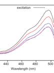
Quantification of Extracellular Ammonium Concentrations and Transporter Activity in Yeast Using AmTrac Fluorescent Sensors

采用AmTrac荧光传感器定量测定酵母细胞的胞外铵浓度和转运蛋白活性

Cindy Ast Cindy Ast
WF Wolf B. Frommer
GG Guido Grossmann
RD Roberto De Michele
11507 Views
Jan 5, 2015
AmTracs are the first example of “activity sensors”, since they report the activity of ammonium transporters by means of fluorescence readout in vivo (De Michele et al., 2013). AmTracs are based on a single fluorescent protein, a circularly permuted GFP (cpGFP), inserted into the cytosolic loop connecting the two pseudosymmetrical halves of the Arabidopsis and yeast plasma membrane ammonium transporters AtAMTs and ScMEP (Figure 1). Recently, FRET-based activity sensors for nitrate and peptide transporters have also been developed (Ho et al., 2014). Since transporter activity directly depends on the availability of substrate, AmTracs measure extracellular ammonium concentrations. Several versions of AmTrac exist, with different fluorescence intensity (FI) responses and affinities for ammonium, and based on different ammonium transporters (AmTrac: AtAMT1;3; AmTrac1;2: At AMT1;2; MepTrac: ScMEP2). Currently, the most useful AmTrac versions are probably AmTrac-GS (bright, with Km of 50 µM) and AmTrac-100 (a high capacity version with Km of 100 µM). The protocol for measuring ammonium concentrations in yeast cells at the fluorimeter is the same for all versions.Figure 1. Model of AmTrac/MepTrac sensors and AMT transport mechanism. We propose that AMT switches between at least two distinct states during transport of ammonium: An outward, open state A and an inward, open state B. The movement of TMH-V (red) and TMH-VI (blue) is transmitted to the connecting loop, affecting the inserted cpGFP (green) and resulting in a change in fluorescence emission.
FWPV 嵌合 MVA的建立

Construction of FWPV Chimaeric MVA

FWPV 嵌合 MVA的建立

KB Karen R. Buttigieg
Michael  A. Skinner Michael A. Skinner
9064 Views
Jan 5, 2015
Construction of chimaeric MVA is a useful tool with which to study gene function of related viruses. The protocol given here describes MVA chimaeras containing genes from Fowlpox virus (FWPV), although this can be applied to DNA derived from other organisms. There are a number of steps required to make the chimaeric MVA: 1) Purification of viral particles; 2) Extraction of DNA from purified viral particles; 3) Assembly of linear recombination templates; 4) Transfection of linear recombination templates; 5) Selection of chimaeric MVA. Note: This procedure uses live virus, and should be conducted using Good Microbiological Practice, in accordance with international and national biocontainment requirements. This procedure also involves Genetic Modification of microorganisms, and appropriate safety approval should be obtained before commencing.

神经科学

神经干细胞、中间神经性前体细胞和未成熟神经元的体外迁移分析

In vitro Migration Assays for Neural Stem Cells, Intermediate Neurogenic Progenitors and Immature Neurons

神经干细胞、中间神经性前体细胞和未成熟神经元的体外迁移分析

JL Julia Ladewig
PK Philipp Koch
Oliver Brüstle Oliver Brüstle
18530 Views
Jan 5, 2015
In the vertebrate central nervous system (CNS), different neural precursor populations such as neural stem cells (NSCs), intermediate neurogenic progenitors (INPs) and immature neurons have to migrate from their places of birth to their location of function. Coordinated migration is mediated by direct cell-cell interactions and by extracellular matrix components, chemoattractants as well as repellents. The migration potential of such populations as well as the responsiveness to chemoattractive compounds can be addressed in isolated cells using in vitro migration assays. Here we describe two migration assays, a matrigel migration assay and a Boyden chamber migration assay, which allow the in vitro assessment of neural migration under defined conditions (Ladewig, Koch and Brüstle, 2014). A matrigel matrix is a soluble basement membrane extract. The major components of matrigel matrix are collagens, laminin and proteoglycans, which provide the substrate for migrating cells. In the matrigel assay migration can be analyzed using a phase contrast microscope. The Boyden chamber assay (Richards and McCullough, 1984) is based on microchemotaxis chambers, which consist of two compartments separated by a membrane with a defined pore size. Cells can be plated in the upper compartment and allowed to migrate through the pores towards the lower compartment, in which a potential chemotactic agent is loaded. Cell migration can be analyzed following fixing and immunohistochemical staining. In principle, the described protocols should be applicable to other cell populations such as endothelial cells or cancer cells using conditions adapted to the individual needs of the specific cell type.
海马神经元细胞培养的锰细胞毒性分析

Manganese Cytotoxicity Assay on Hippocampal Neuronal Cell Culture

海马神经元细胞培养的锰细胞毒性分析

Alexia Daoust Alexia Daoust
YS Yasmina Saoudi
JB Jacques Brocard
NC Nora Collomb
Cécile Batandier Cécile Batandier
MB Mariano Bisbal
MS Murielle Salomé
AA Annie Andrieux
SB Sylvain Bohic
EB Emmanuel L. Barbier
9357 Views
Jan 5, 2015
Compared to an in vivo experiment, neuronal cell cultures are immediately accessible to observation and manipulation. In this protocol, we describe a technique to evaluate the cytotoxicity of a metal, manganese (Mn2+), on hippocampal neuronal cell cultures. Interestingly, this protocol is easily adaptable to any type of primary culture (e.g., cortical neurons) and any type of toxic compound (e.g., chemical product).This protocol is similar to "Neuron-enriched Cultures (Method 2)" protocol (Gao, 2011).

植物科学

浮萍根部分泌物的采集

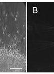
Collection of Root Exudate from Duckweed

浮萍根部分泌物的采集

Yufang Lu Yufang Lu
LS Li Sun
Weiming Shi Weiming Shi
13981 Views
Jan 5, 2015
Root exudates play an important role in rhizosphere interactions between plants and microorganisms. However, collection of chemicals from plant root system is difficult due to their low concentrations and high level of contaminants in growth media. The continuous collection method has been described in several terrestrial plants over the past 30 years (Tang and Young, 1982; Kong et al., 2004). Here, we describe a protocol for the collection of sterile root exudate from a floating aquatic plant, duckweed.
胚胎油体等球状体大小、分布的成像和定量测定

Imaging and Quantitative Analysis of Size and Distribution of Spherical Bodies, e.g. Embryonic Oil Bodies

胚胎油体等球状体大小、分布的成像和定量测定

Martine Miquel Martine Miquel
GT Ghassen Trigui
AT Alain Trubuil
Bertrand Dubreucq Bertrand Dubreucq
8688 Views
Jan 5, 2015
Oil bodies (OBs) are seed-specific lipid storage organelles that allow the accumulation of neutral lipids that sustain plantlet development after the onset of germination. Using fluorescent dyes and confocal microscopy, we monitored the dynamics of OBs in living Arabidopsis (Arabidopsis thaliana) embryos at different stages of development (Miquel et al., 2014). Image acquisition was followed by a detailed statistical analysis of OB size and distribution during seed development in the four dimensions (x, y, z, and t).