往期刊物2014

卷册: 4, 期号: 13

left

Dec

20

Dec

5

Nov

20

Nov

5

Oct

20

Oct

5

Sep

20

Sep

5

Aug

20

Aug

5

Jul

20

Jul

5

Jun

20

Jun

5

May

20

May

5

Apr

20

Apr

5

Mar

20

Mar

5

Feb

20

Feb

5

Jan

20

Jan

5

right

免疫学

鼠体内 CD8+ T淋巴细胞杀菌分析

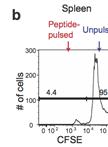
Murine in vivo CD8+ T Cell Killing Assay

鼠体内 CD8+ T淋巴细胞杀菌分析

MK Myoungjoo V. Kim
WO Weiming Ouyang
WL Will Liao
MZ Michael Q. Zhang
ML Ming O. Li
19075 Views
Jul 5, 2014
Antigen-specific killing ability of effector CD8+ T cells is critical for protective immunity against infection. Here, we describe in vivo cytotoxic T cell assay to examine effector function of antigen-specific CD8+ T cells. Mice infected with Listeria monocytogenes (L. monocytogenes) expressing chicken ovalbumin as a model antigen mount ovalbumin-specific CD8+ T cell responses. Effector CD8+ T cell function in vivo is determined by mixed transfer of OVA peptide-pulsed target cells with control target cells into the previously immunized mice. Difference in CFSE expression levels clearly marks two distinct populations: Antigen-pulsed target cells-CFSElow vs. unpulsed target cells-CFSEhi. The frequencies between antigen-pulsed target cells and control target cells are used as readouts of antigen-specific killing.
吞噬溶酶体的胞内转运分析

Phagolysosomal Trafficking Assay

吞噬溶酶体的胞内转运分析

Alka  Mehra Alka Mehra
15914 Views
Jul 5, 2014
Phagolysosomal trafficking is an important innate defense pathway that clears microbes by delivering them to lysosomes, the degradative compartment of the cell. Mycobacterium tuberculosis (Mtb), the causative agent of tuberculosis, subverts this host defense mechanism by arresting maturation of the phagosome. The ability of Mtb to arrest its delivery to the lysosome can be demonstrated by the prolonged co-localization of bacteria containing phagosomes/vacuole with early phagosomal markers [such as, Ras- related proteins in the brain 5 (Rab5) and Transferrin receptor (TfR)], and a failure to acquire late phagosomal and lysosomal markers (such as Rab7 and LAMP1) (Deretic and Fratti, 1999, Mehra et al., 2013). Here, a protocol is outlined for infection of macrophages with mycobacterial species like pathogenic Mtb, vaccine strain Mycobacterium bovis- bacillus Calmatte- Guérin (BCG) and rapidly dividing non-pathogenic Mycobacterium smegmatis (Msmeg), followed by indirect-immunofluorescence microscopy to visualize host vacuolar markers. Thereafter, automated quantification of degree of co-localization between mycobacteria and host vacuolar markers like TfR and LAMP1 is done by processing the binary images of bacteria using mathematical tools. This results in quantification of the mean fluorescence intensity (MFI) of these host markers directly around the bacteria/bacterial clusters with increased sensitivity relative to when done manually. By manipulating host or pathogen, this assay can be used to evaluate host or bacterial determinants of intracellular trafficking. The basic method can be applied to studying trafficking of other bacteria or particles like beads, although the kinetics of infection and phagosome maturation will depend upon the phagocytic cargo. The mathematical analysis tools are available in many standard imaging analysis programs. However, any adaption for similar analysis should be confirmed by the individual user with their imaging and analysis platform.
鼠源记忆性T淋巴细胞的体外分化

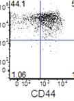
Murine in vitro Memory T Cell Differentiation

鼠源记忆性T淋巴细胞的体外分化

MK Myoungjoo V. Kim
WO Weiming Ouyang
WL Will Liao
MZ Michael Q. Zhang
ML Ming O. Li
14651 Views
Jul 5, 2014
Upon pathogen encounter, naïve CD8+ T cells are primed and undergo massive clonal expansion. A fraction of effector CD8+ T cells remains during the contraction phase and differentiate into memory T cells critical for mounting robust recall responses in response to secondary infection. Low frequency of memory T cells in vivo is a major obstacle to investigate their functional aspects including migration capacity and genetic regulation. Here, we describe detailed protocol for memory T cell differentiation developed by von Andrian’s group to generate large number of CD44hiCD62Lhi antigen-specific memory T cells in vitro.
悬浮细胞的脉冲追踪

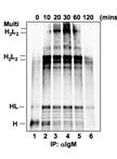
Pulse Chase of Suspension Cells

悬浮细胞的脉冲追踪

LW Lai-Yee Wong
QiMing  Liang QiMing Liang
Kevin  Brulois Kevin Brulois
Jae  Jung Jae Jung
12654 Views
Jul 5, 2014
Pulse-chase method is a powerful technique used to follow the dynamics of proteins over a period of time. The expression level, processing, transport, secretion or half-life of proteins can be tracked by metabolically labeling the cells, such as with radiolabeled amino acids (pulse step). This protocol describes the condition used to study the folding and disulfide bond formation of immunoglobulin in suspension cells. With some minor modifications, this protocol can be adapted to study the degradation rate or the secretion of target proteins.

微生物学

分枝杆菌中利福平耐药性突变频率的测定和突变频谱分析

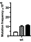
Determination of Rifampicin-resistance Mutation Frequency and Analysis of Mutation Spectra in Mycobacteria

分枝杆菌中利福平耐药性突变频率的测定和突变频谱分析

LL Liang-Dong Lyu
GZ Guo-Ping Zhao
11886 Views
Jul 5, 2014
Understanding the genetic safeguarding mechanism of Mycobacterium tuberculosis (Mtb) may help us to explain i), how Mtb survive the genetic assaults elicited by both reactive oxygen species (ROS) and reactive nitrogen species (RNS) produced by host macrophages and ii), why some strains of Mtb, e.g., Mtb strains from East Asian lineage and Beijing sublineage, exhibit high mutation rate and are more likely to acquire drug resistant mutations (e.g., rifampicin-resistance mutation) during infection. Mutation frequency analysis is a basic methods to study the genetic safeguarding mechanism. Moreover, to study the molecular mechanism of mutation, it is necessary to analyse the mutation spectrum (For example, oxidized cytosine may induce CG to TA mutation.). This protocol describes a method to determine the mutation frequency and understand the mutation spectrum in both Mycobacterium smegmatis (Msm) and Mtb.

神经科学

雏鸡胚胎的开放式制备和合缝轴突的DiI标记

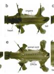
Open-book Preparations from Chick Embryos and DiI Labeling of Commissural Axons

雏鸡胚胎的开放式制备和合缝轴突的DiI标记

Nicole  H.  Wilson Nicole H. Wilson
Esther  T.  Stoeckli Esther T. Stoeckli
12685 Views
Jul 5, 2014
Successful neural circuit formation relies on the accurate navigation of axons towards their targets during development. Axons are guided by a combination of short-range and long-range, attractive and repulsive cues. The commissural axons of the developing spinal cord have provided an informative in vivo model for the identification of multiple axon guidance molecules and mechanisms. These axons extend ventrally from the dorsal spinal cord and cross the midline at the floor plate, before making a sharp rostral turn towards the head. This simple trajectory has facilitated the identification of many axon guidance molecules, because perturbation of the stereotypical guidance decisions as a result of genetic manipulations can be easily identified. The open-book assay is a method to assess the trajectory of spinal commissural axons. The spinal cord is dissected out, opened at the roof plate and pinned flat. Punctate injections of the lipophilic fluorescent dye, DiI, are used to trace commissural axon trajectories prior to microscopy and analysis.

植物科学

采用细胞计数器插件分析植株叶片细胞大小、密度和数量以及表皮毛数量

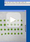
Analyses of Plant Leaf Cell Size, Density and Number, as Well as Trichome Number Using Cell Counter Plugin

采用细胞计数器插件分析植株叶片细胞大小、密度和数量以及表皮毛数量

YC Yan Cheng
LC Ling Cao
SW Sheng Wang
YL Yongpeng Li
HW Hong Wang
YZ Yongming Zhou
26520 Views
Jul 5, 2014
An Arabidopsis leaf blade is composed of many layers that are sandwiched between two layers of tough skin cells (called the epidermis). Four layers (adaxial epidermis, palisade layer, spongy mesophyll and abaxial epidermis) contain specialized cells. Here we describe a quick and simple method for analyzing the size, number and density of different types of cells in an Arabidopsis leaf blade. This method would be of interest to people who would like to investigate cell size and number changs in different cell layers in leaves or leaf-like organs without having to dissect the samples.
拟南芥嫁接实验

Grafting Arabidopsis

拟南芥嫁接实验

TA Tonni Grube Andersen
DL Dacheng Liang
BH Barbara Ann Halkier
RW Rosemary White
16812 Views
Jul 5, 2014
In Arabidopsis thaliana, hypocotyl micrografting has been used to investigate transport of flowering signals, mobile silencing signals and other peptides, proteins and secondary compounds. The effects of transported signals on target tissues require that a good vascular connection is re-established across the graft junction between the cut hypocotyls (stumps) of the root (rootstock) and shoot (scion) tissues. We outline here a method that requires only that the cut stumps be placed in close proximity, so that they touch, followed by 3-5 days of undisturbed recovery time during which the grafts are allowed to dry out somewhat. This method is quick, easy to monitor and has up to 90% success rate.
优化用于Illumina测序的植物序列捕获技术

Plant Sequence Capture Optimised for Illumina Sequencing

优化用于Illumina测序的植物序列捕获技术

Axel  Himmelbach Axel Himmelbach
Manuela  Knauft Manuela Knauft
NS Nils Stein
15667 Views
Jul 5, 2014
Plant Sequence Capture is used for targeted resequencing of whole exomes (all exons of a genome) of complex genomes e.g. barley and its relatives (Mascher et al., 2013). Sequencing and computing costs are significantly reduced since only the greatly enriched and gene-coding part of the barley genome is targeted, that corresponds to only 1-2% of the entire genome. Thus, applications such as genetic diversity studies and the isolation of single genes (“cloning-by-sequencing”) are greatly facilitated. Here, a protocol is provided describing the construction of shotgun DNA libraries from genomic barley DNA for sequencing on the Illumina HiSeq/MiSeq systems. The shotgun DNA sequencing libraries are hybridized to an oligonucleotide pool (Exome Library) encompassing the whole exome of barley. The Exome Library is provided as a liquid array containing biotinylated probes (Roche/NimbleGen). Subsequently, genomic shotgun DNA fragments hybridized to the Exome Library are affinity-purified using streptavidin coated magnetic beads. The captured library is PCR-amplified and sequenced using high-throughput short read sequencing-by-synthesis.
细胞分裂素分析:样本制备和定量测定

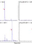
Cytokinin Analysis: Sample Preparation and Quantification

细胞分裂素分析:样本制备和定量测定

Martin  Schäfer Martin Schäfer
MR Michael Reichelt
IB Ian T. Baldwin
SM Stefan Meldau
15624 Views
Jul 5, 2014
Cytokinins are a group of phytohormones discovered about half a decade ago by Miller et al. (1955) and Skoog et al. (1965). Since then they were found to participate in many plant physiological processes, including the regulation of the source/sink transitions, plant growth and organ development, responses to environmental conditions such as light, nutrient and water availability and biotic interactions with mutualists, pathogens and herbivores (Werner and Schmülling, 2009; Giron et al., 2013). To aid the quantification of cytokinins for analyzing their changes after environmental stress conditions, we developed this cytokinin extraction and analysis method. This protocol is based on the cytokinin extraction with an acidic methanol-water solution and purification with a mixed-mode solid phase extraction procedure described by Dobrev and Kamı́nek (2002) and the modifications of Kojima et al. (2009). The protocol was successfully used to verify cytokinin overproduction in transgenic Nicotiana attenuata plants expressing the cytokinin biosynthesis gene Tumor morphology root (Tmr) from Agrobacterium tumefaciens under the control of the chemical inducible expression system pOp6/LhGR in the glasshouse and under field conditions (Schäfer et al., 2013) to study the role of cytokinins in plant-herbivore interactions.
种皮渗透性测试:四唑四氮唑蓝渗透分析

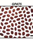
Seed Coat Permeability Test: Tetrazolium Penetration Assay

种皮渗透性测试:四唑四氮唑蓝渗透分析

Sollapura  J.  Vishwanath Sollapura J. Vishwanath
Frédéric  Domergue Frédéric Domergue
OR Owen Rowland
14174 Views
Jul 5, 2014
Seed coat permeability is important to study as it plays significant roles in seed dormancy, germination, and protection from pathogens. Here we describe a commonly used seed coat permeability test known as the tetrazolium penetration assay with a method to quantify the levels of permeability. Tetrazolium red is a cationic dye that is widely used in seed viability testing. Tetrazolium salts are amphipathic cations, which, after penetrating the dead cells of the seed coat, are reduced to red-colored insoluble precipitates made up of formazans by active dehydrogenases (NADH-dependent reductases) in the embryo of seeds (Berridge et al., 1996). The intensity of red coloration is directly proportional to the permeability of the seeds. The quantification involves extraction of formazans from the incubated seeds and spectrophotometric determination of absorbance of formazan extracts at 485 nm. Note: This protocol is optimized for testing Arabidopsis thaliana seeds.
大麦幼苗中磷分布的放射性自显影

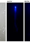
Autoradiography of Pi Distribution in Barley Seedlings

大麦幼苗中磷分布的放射性自显影

Satomi  Kanno Satomi Kanno
Yuko  Kurita Yuko Kurita
Miwa  Ohnishi Miwa Ohnishi
Tetsuro  Mimura Tetsuro Mimura
9093 Views
Jul 5, 2014
Phosphorus-32 and Phosphorus-33 are radioisotopes of phosphorus. These isotopes are used to trace ionic phosphorus and phosphorus compounds. This protocol is used to follow the movement of inorganic phosphate (PO43-) from a leaf tip to the rest of the plant.
提取木材中的非结构性碳和纤维素用于放射性碳素分析

Extraction of Nonstructural Carbon and Cellulose from Wood for Radiocarbon Analysis

提取木材中的非结构性碳和纤维素用于放射性碳素分析

CC Claudia I. Czimczik
ST Susan E. Trumbore
XX Xiaomei Xu
MC Mariah S. Carbone
AR Andrew D. Richardson
8740 Views
Jul 5, 2014
This method aims at isolating nonstructural organic carbon (NSC) pools, i.e. soluble sugars and starch, from wood for radiocarbon (14C) analysis at natural abundance levels (≤1 ppt). Pools are operationally defined to 1) physically isolate pools - prohibiting the use of destructive methods, such as compound-specific enzyme digestion, and 2) minimize possible contamination with extraneous carbon form organic solvents.
叶片切片的离子提取

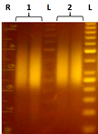
Extraction of Ions from Leaf Sections

叶片切片的离子提取

Yuko  Kurita Yuko Kurita
Satomi  Kanno Satomi Kanno
Miwa  Ohnishi Miwa Ohnishi
Tetsuro  Mimura Tetsuro Mimura
7196 Views
Jul 5, 2014
The concentration of ions in plant cells and tissues is an important factor to determine their functions and conditions. Here, we describe the method to extract ions from leaf sections for measurements with an ion chromatogram. This method is available for not only barley but also other plant species.