Next-generation expansion microscopy: 20× magnification in one step
Speaker: Shiwei Wang
Online live: May 20, 2025 11:00 AM ET | 8:00 AM PT Posted: May 26, 2025 Views: 11533
 WEBINAR SERIES
Abstract
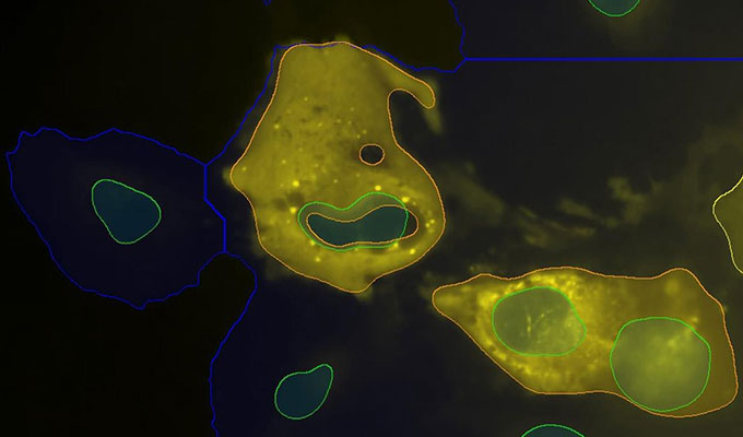
Expansion microscopy (ExM) has revolutionized biological imaging by enabling nanoscale-resolution visualization on conventional microscopes. Traditional ExM methods have been limited to ~4–10× expansion per step or require iterative processes to achieve higher magnifications (~15–20×). In this webinar, we will introduce 20ExM, a novel single-shot ExM protocol that achieves ~20× expansion in a single step, providing <20 nm resolution. This innovation simplifies high-resolution imaging while supporting postexpansion biomolecular staining in brain tissue. The discussion will cover the principles of ExM, the advantages of 20ExM over iterative expansion, and its potential applications in neuroscience, cell biology, and beyond.
Highlights
- Fundamentals of expansion microscopy (ExM) and its impact on high-resolution imaging.
- 20ExM: A single-shot expansion protocol achieving ~20× linear expansion.
- Post-expansion antibody staining for brain tissue and its implications.
- Applications of 20ExM in neuroscience and cell biology and comparison with previous ExM approaches.
Speaker

Shiwei Wang, B.A.
Ph.D. candidate, Massachusetts Institute of Technology
Shiwei Wang is a Ph.D. candidate in Chemical Biology at the Massachusetts Institute of Technology (MIT), where he develops innovative tools for neu...
View more ![]()
Keywords
Expansion microscopy (ExM), Super-resolution imaging techniques, Nanoscale visualization, Brain tissue imaging, Biomolecular labeling, Confocal microscopy
References
Wang, S., Shin, T.W., Yoder, H.B. et al. Single-shot 20-fold expansion microscopy. Nat Methods 21, 2128–2134 (2024). https://doi.org/10.1038/s41592-024-02454-9
Do you have a question about this webinar?
Post your question, and we'll invite the webinar speaker to respond. You're welcome to join the discussion by answering or commenting on questions ( Note: Not all questions, especially those not directly relevant to the webinar topic, may be answered by the speaker. ).
![]()
+
36 Q&A
What happens to structures that are known and regulated to occupy a specific amount of space?
What advancements enable 20× magnification in a single step in next-generation expansion microscopy?
 Jun 18, 2026
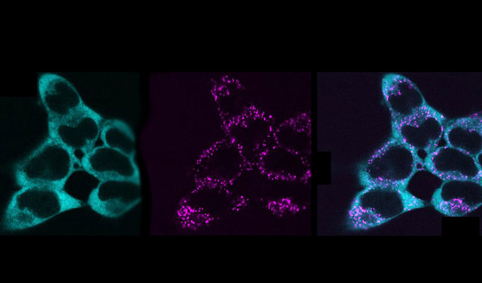 May 27, 2026
 Apr 28, 2026