Label retention expansion microscopy (LR-ExM) with Aydin denoising
Speakers: Xiang Zhao and Ahmet Can Solak Moderator: Xiaoyu Shi
Online live: Nov 08, 2022 12:00 PM ET | 9:00 AM PT Posted: Nov 21, 2022 Views: 3304

 WEBINAR SERIES
Abstract
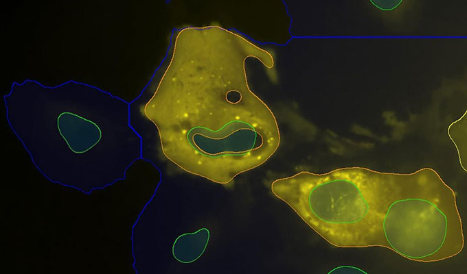
Expansion microscopy (ExM) developed in the recent decade has enabled conventional confocal microscopy to be used for super-resolution imaging by physically expanding the sample within swellable hydrogel. Since its invention, ExM has been tested with many different types of biological samples. The main challenge addressed frequently is the fluorescence signal loss during polymerization and digestion of ExM protocols. The recently developed label-retention ExM has solved the problem with trifunctional anchors. LR-ExM has enabled high-efficiency labeling and prevented the signaling loss, but it has also increased the signaling noises meanwhile. Here we have established the protocol for applying LR-ExM with zebrafish cryosections at first. Together with Aydin denoising, we have optimized the imaging data set and obtained high quality super-resolution immunofluorescent images. Leverage on both LR-ExM with Aydin denoising tools, we have identified that cytosolic Par-3 is associated with Dynein complex and Dld on Rab11 endosomes in the mitotic radial glia cells in zebrafish forebrain.
Speakers
Xiang Zhao, Ph.D.
Research Scientist, Chan Zuckerberg Biohub / University of California San Francisco
Xiang Zhao is a neuroscientist enthusiastic about the gene function and regulation mechanism in the development of the central nervous system. Xian...
View more ![]()
Ahmet Can Solak, BSc.
Software Engineer, Chan Zuckerberg Biohub
Ahmet Can Solak, got his BSc. on Electrical and Electronics Engineering in 2018 from Koc University in Istanbul, Turkey. He joined the Loic Royer’s...
View more ![]()
Moderator

Xiaoyu Shi, Ph.D.
Assistant Professor, University of California
Xiaoyu Shi is an Assistant Professor in the Department of Developmental and Cell Biology and the Department of Chemistry at the University of Calif...
View more ![]()
Keywords
Expansion microscopy, Aydin imaging denoising, Par-3, Endosome, Asymmetric cell division
References
Zhao, X., Garcia, J.Q., Royer, L.A., and Guo, S. (2022) Colocalization Analysis for Cryosectioned and Immunostained Tissue Samples with or without Label Retention Expansion Microscopy (LR-ExM) by JACoP. Bio-protocol 12(5): e4336. PMCID: PMC8918214.
Do you have a question about this webinar?
Post your question, and we'll invite the webinar speaker to respond. You're welcome to join the discussion by answering or commenting on questions ( Note: Not all questions, especially those not directly relevant to the webinar topic, may be answered by the speaker. ).
![]()
+
22 Q&A
How efficient the technique is to work with primary and fluorophore tagged secondary antibodies?

 Jun 18, 2026
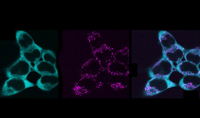 May 27, 2026
 Apr 28, 2026