- Protocols

- Articles and Issues
- For Authors
- About
- Become a Reviewer

pERK Detection Assays Using the Surefire AlphaScreen® Kit (TGR Biosciences and PerkinElmer)
Published: Vol 3, Iss 13, Jul 5, 2013 DOI: 10.21769/BioProtoc.806 Views: 10613
Reviewed by: Cheng Zhang
Original research article
The authors used this protocol in:

Nov 2012

Protocol Collections
Comprehensive collections of detailed, peer-reviewed protocols focusing on specific topics


Related protocols
Abstract
Extracellular signal-regulated kinase 1 and 2 (ERK1/2) are serine/threonine protein kinases that are phosphorylated on Thr202/Tyr204 (ERK1) and Thr185/Tyr187 (ERK2). Phosphorylation of ERK1/2 (pERK1/2) arises from multiple stimuli, resulting in physiological responses that include cell growth, proliferation and differentiation. This protocol has been optimized for the detection of ligand-mediated pERK1/2 in adherent immortal cell lines expressing G protein-coupled receptors (GPCRs).
Materials and Reagents
- Dulbecco’s modified eagle medium (DMEM) (Life Technologies, Gibco®, catalog number: 11995-065 )
- Bovine serum albumin (BSA) (Sigma-Aldrich, catalog number: A7906 )
- SureFire® Reagents (Includes Lysis, Activation and Reaction buffers) (TGR BioSciences, catalog number: TGRES500 )
- AlphaScreen® General IgG (Protein A) detection kit (PerkinElmer, catalog number: 6760617 )
- White ProxiPlate 384-well microplate (PerkinElmer, catalog number: 6008280 )
- TopSeal (PerkinElmer, catalog number: 6005250 )
- NaCl
- KCl
- Na2HPO4
- KH2PO4
- Phosphate buffered saline (PBS) (see Recipes)
- Detection buffer (see Recipes)
Equipment
- Fusion-α plate reader or Envision plate reader with appropriate Alphascreen detection modules (PerkinElmer)
- Sterile 96-well clear flat bottom plates (BD Biosciences, Falcon®, catalog number: 353072 )
- Humidified incubator
- Multichannel pipettes
- Micropipettes
- Orbital shaker
Procedure
Notes:
1) Cells can either be stably or transiently expressing receptor of interest.
2) It is recommended to first perform a timecourse analysis to determine the time at which ligand-mediated pERK1/2 is maximal. For this, follow the same protocolusing a single concentration of ligand (recommended concentration 100x Kd).
Recommended initial timecourse (min): 90, 60, 45, 30, 15, 10, 8, 6, 4, 2, 1, 0.
3) Subsequent timecourses can then be refined to determine the precise time at which maximum ligand-induced pERK1/2 occurs.
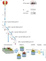
I. Cell preparation
Seed cells in suitable nutrient media (e.g. DMEM, 10% FBS, no antibiotics) into a sterile 96-well plate and incubate in a humidified environment at 37 °C, 5% CO2 to be ~90% confluent the following day (~24 h).
Note: Optimization for cell number depending on the cell line used will be necessary (we suggest a starting range of 10,000-50,000 cells per well). Recommended density for CHO FlpIN cells is 30,000 cells/ well.
II. Stimulation
- The day following seeding, aspirate nutrient media, rinse once with 100 μl PBS, and replace with 90 μl prewarmed DMEM (no FBS).
- Incubate in a humidified environment at 37 °C, 5% CO2 for a minimum of 4 h (recommended 6 h, up to overnight (O/N)).
- Prepare serial dilutions of ligands at 10x final concentration in DMEM, enough for 10 μl/well, to be performed in duplicate (minimum), and enough for the number of timepoints if doing a timecourse.
Note: Concentration range to use will depend on ligand affinity for receptor. For initial timecourse test, select a concentration 100x Kd of ligand and a DMEM control. The concentrations can then be refined in subsequent experiments. If using a peptide or ‘sticky’ ligand, prepare serial dilutions in DMEM with 0.1% BSA.
- Prepare a suitable concentration of FBS in DMEM, enough for 10 μl/well, to be performed in duplicate (minimum), and enough for the number of timepoints if doing a timecourse.
Note: This is the internal control for the experiment – FBS promotes pERK1/2. Recommended final concentration of FBS in a CHO FlpIN cell line is 3-10%.
- Following preincubation in DMEM, add 10 μl of 10x prepared ligands or FBS to cells for a total volume of 100 μl, 1x final concentration.
Note: For initial timecourse, begin at 90 min, and add ligand or FBS to cells at each timepoint until time 0 (no addition). For concentration response, add ligand or FBS at time of maximal induced pERK1/2 as determined through timecourse.
- After completion of stimulation, rapidly remove ligand-containing media from cells.
Note: Depending on the cell type, this may involve flicking or gentle aspiration.
- Add 50 μl 1x Surefire® Lysis buffer.
Note: Optimization for lysis volume will be necessary, and depends on the cell type, expression level of the receptor and efficiency of coupling to pERK1/2 pathways. Recommended starting lysis volume in a CHO FlpIN cell line is 30-100 μl.
- Incubate lysates at room temperature (RT) for 5-10 min on an orbital shaker.
III. Detection
- In reduced lighting conditions, prepare detection buffer.
- Transfer 5 μl of cell lysate from each well to a 384-well ProxiPlate.
- In reduced lighting conditions, add 8.5 μl Detection buffer to each sample.
- Seal the plate with TopSeal and wrap in foil.
Note: Small volumes are subject to evaporation, TopSeal is essential.
- Incubate at RT for 2 h or 37 °C for 1 h in reduced lighting conditions.
Note: If incubating at 37 °C, ensure the plate has returned to RT before measuring luminescence (~15 min at RT following 37 °C incubation should suffice). Detection beads are temperature sensitive.
- Analyse luminescence on a Fusion-α or Envision plate reader using standard α-screen settings.
IV. Data analysis
Data should be normalized to the response elicited by the FBS control.
Recipes
- Phosphate Buffered Saline (PBS)
137 mM NaCl, 2.7 mM KCl, 10 mM Na2HPO4, 1.8 mM KH2PO4, pH 7.4.
- Detection buffer
85.0% SureFire® reaction buffer
14.2% SureFire® activation buffer*
0.4% Acceptor beads
0.4% Donor beads
* Activation buffer should be stored at 4 °C, however, precipitation will occur at this temperature. Before use, heat to 37 °C to ensure all is dissolved.
Prepare Detection buffer immediately before use. Discard unused detection buffer. Mix detection buffer gently. Do not vortex.
Additional note: Lysates may be stored at -20 °C and pERK1/2 detected at a later time, but no longer than 2 weeks following stimulation.
References
- Koole, C., Wootten, D., Simms, J., Valant, C., Sridhar, R., Woodman, O. L., Miller, L. J., Summers, R. J., Christopoulos, A. and Sexton, P. M. (2010). Allosteric ligands of the glucagon-like peptide 1 receptor (GLP-1R) differentially modulate endogenous and exogenous peptide responses in a pathway-selective manner: implications for drug screening. Mol Pharmacol 78(3): 456-465.
Article Information
Copyright
© 2013 The Authors; exclusive licensee Bio-protocol LLC.
How to cite
Koole, C., Wootten, D. and Sexton, P. M. (2013). pERK Detection Assays Using the Surefire AlphaScreen® Kit (TGR Biosciences and PerkinElmer). Bio-protocol 3(13): e806. DOI: 10.21769/BioProtoc.806.
Category
Cell Biology > Cell signaling > Phosphorylation
Biochemistry > Protein > Modification
Biochemistry > Protein > Interaction > Protein-ligand interaction
Do you have any questions about this protocol?
Post your question to gather feedback from the community. We will also invite the authors of this article to respond.