- Protocols

- Articles and Issues
- For Authors
- About
- Become a Reviewer

Preparation of Teased Nerve Fibers from Rat Sciatic Nerve
Published: Vol 7, Iss 19, Oct 5, 2017 DOI: 10.21769/BioProtoc.2572 Views: 12324
Reviewed by: Alexandros KokotosAnonymous reviewer(s)
Original research article
The authors used this protocol in:
Mar 2017
Advertisement


Protocol Collections
Comprehensive collections of detailed, peer-reviewed protocols focusing on specific topics


Abstract
Compared to tissue sectioning techniques, the technique of teasing single nerve fibers provides a better way to understand the structures of myelin sheaths and axons of the peripheral myelinated nerves. This protocol describes a method for preparation of teased single nerve fibers from rat sciatic nerve. In this protocol, fixed nerves are teased into single individual fibers and arranged onto adhesion microscope slides for further immuno-staining.
Keywords: Peripheral nerveBackground
Schwann cells in the peripheral nervous system wrap around axons to form insulated myelin sheaths that allow the rapid conduction of action potentials. The remarkable multi-layered myelin sheath consists of elaborate structures including compact sheath, Schmidt-Lanterman incisures, Cajal bands, inner and outer mesaxons, as well as the structures in the paranodal region. To elucidate the normal and abnormal structures of myelinated fibers of peripheral nerves, teased nerve fibers are required. Methods of teasing fibers have been widely applied in studies on peripheral nerves of human and rodents. In this protocol, we describe a method of teasing peripheral nerves into single fibers for further morphological studies on the axons or myelin of peripheral nerves.
Materials and Reagents
- Adhesion microscope slides (CITOTEST LABWARE MANUFACTURING, catalog number: 80312-3161-16 )
Note: These slides with special treatment process that electrostatically adheres tissue to the glass without the need for adhesives or protein coatings.
- Cover slips (CITOTEST LABWARE MANUFACTURING, catalog number: 80342-1130 )
- Cell culture dish (100 x 10 mm) (Corning, catalog number: 430167 )
- Animal: 4-month-old Sprague Dawley (SD) rat
- Chloral hydrate (Sinopharm Chemical Reagent, catalog number: 30037517 )
- Sodium chloride (NaCl) (Guangdong Guanghua Sci-Tech, catalog number: 1.01307.040 )
- Paraformaldehyde (PFA) (Guangdong Guanghua Sci-Tech, catalog number: 1.17767.014 )
- Phosphate-buffered saline (PBS) (Beyotime Biotechnology, catalog number: C0221A )
- Triton X-100 (Sigma-Aldrich, catalog number: V900502 )
- Gelatin (Sigma-Aldrich, catalog number: G7041 )
- Anti-S100 Protein antibody, clone 15E2E2, produced in mouse (S100) (Merck, catalog number: MAB079-1 )
- Anti-Neurofilament 200 antibody produced in rabbit (NF) (Sigma-Aldrich, catalog number: N4142 )
- Alexa Fluor® 488 goat anti-mouse IgG (H+L) (Thermo Fisher Scientific, InvitrogenTM, catalog number: A-11001 )
- Alexa Fluor® 568 goat anti-rabbit IgG (H+L) (Thermo Fisher Scientific, InvitrogenTM, catalog number: A-11011 )
- TWEEN® 20 (Sigma-Aldrich, catalog number: P1379 )
- 4,6-Diamidino-2-phenylindole (DAPI) (Sigma-Aldrich, catalog number: D9542 )
- Mounting medium for fluorescence (Vector Laboratories, catalog number: H-1000 )
- 10% chloral hydrate (see Recipes)
- 0.9% NaCl (see Recipes)
- 4% PFA (see Recipes)
- 0.1% Triton X-100 (see Recipes)
- Blocking buffer (see Recipes)
- PBST (see Recipes)
- 1,000x DAPI (see Recipes)
Equipment
- Dissecting scissors and forceps (see Figure 1A)
- Perfusion pump (Longer, catalog number: BT300-1F )
- Spring scissors (66 Vision Tech, catalog number: 54053B ) (see Figure 2A)
- Fine forceps (Fine Science Tools, Dumont, model: #5SF, catalog number: 11252-00 , see Figure 2A)
- Black wet chamber (Leibusi, catalog number: 340012 )
Note: The wet chamber (Leibusi, catalog number: 340012 ) was made by a local workshop.
- Stereomicroscope (Olympus, catalog number: SZ61 )
Procedure
- Anaesthetize a rat with an intraperitoneal injection of 10% chloral hydrate (0.3 ml/100 g) (see Recipes).
Note: Animal use was approved by the Southern Medical University Animal Care and Use Committee and the experimental procedure was performed in accordance with the guidelines for the ethical treatment of animals. All efforts were made to minimize animal sacrifice and suffering.
- With big scissors and forceps, expose the heart by cutting the thoracic cavity open.
- Using a perfusion pump, perfuse the rat with cold 0.9% NaCl (see Recipes) by transcardial perfusion to clear the blood in the body.
- Perfuse the rat with cold 4% PFA (see Recipes) to fix the rat till it becomes stiff.
- With big scissors and forceps, cut the skin to expose the biceps femoris and the intermuscular septum (see Figure 1B).
- With small scissors and forceps, separate the biceps femoris along the intermuscular septum to expose the sciatic nerve (see Figure 1C).

Figure 1. Expose the sciatic nerve. A. Scissors and forceps used to dissect sciatic nerves; B. The biceps femoris (BF) and the intermuscular septum (IS) are shown after removal of the skin. C. Sciatic nerve is exposed after dissecting the biceps femoris. C’. A magnified image of C shows the sciatic nerve before removal of it from the rat.
- Use small scissors to cut the nerve at the knee and the sciatic notch, obtain the nerve and store in 4% PFA at 4 °C overnight for post-fixation.
- Use spring scissors to cut a nerve segment of 3-5 mm from the fixed nerve, and place the segment in a dish filled with PBS at room temperature (RT) (see Figure 2B).
- Use fine forceps to remove the epineurium wrapping the nerve under a stereomicroscope (see Figure 2C).
Note: The epineurium is the outermost layer of dense irregular connective tissue surrounding the nerve. It appears more transparent than the nerve fasciculus after the fixation. - Transfer the epineurium-free nerve onto an adhesion slide immersed in PBS in a dish (see Figure 2D).
- Use fine forceps to tease the nerve into parts under the stereomicroscope and carefully isolate the single individual fibers (see Figure 2E).
Note: While teasing the nerve into parts, some fibers will dissociate from the nerve fasciculus individually or in small groups. Then isolate the dissociated fibers from the fasciculus and carefully tease them into single individual fibers without breaking them. Isolated fibers floating in PBS appear white in color and semi-transparent. - Use fine forceps to carefully brush the single individual nerve fibers onto the adhesion microscope slide (see Figure 2F).
Note: Good adhesion of fibers to slides is essential in this step, and make sure the fibers are well adhered to the slides. The whole fiber attaching to the slide without any detachment suggests good adherence.

Figure 2. Step-by-step procedures of teasing individual fibers. A. Spring scissors and forceps used to tease nerve; B. A nerve segment before the removal of epineurium; C. Nerve epineurium (arrow) is half stripped off from the nerve and the epineurium-free nerve fasciculus (arrowhead). D. Nerve fasciculus immersed in PBS on an adhesion slide in a dish under a stereomicroscope; E. Nerve fasciculus with single individual fibers; F. Straight single individual nerve fibers adhering to the slide.
- Lift the slide out of PBS and absorb the liquid left on the slide with absorbent paper.
- Air-dry the adhering fibers and store at 4 °C overnight for stronger adhesion.
- Immuno-stain the teased fibers (steps 16-26) in the following days as soon as possible for further analysis.
Note: Store slides at -80 °C for long-term storage. However, immunofluorescent staining performed in the next day usually gets the most guaranteed results.
- Wash the fibers on the slides 3 times (10 min each) with PBS.
- Incubate the fibers with 0.1% Triton X-100 (see Recipes) in PBS for 30 min to penetrate the membrane.
- Incubate the fibers with blocking buffer (see Recipes) for 1 h at RT.
- Incubate the fibers with primary antibodies overnight at 4 °C.
Note: Primary antibodies of anti-S100 (1:400, diluted in blocking buffer) to show Schwann cell cytoplasm) and anti-NF (1:800, diluted in blocking buffer) to show neuron axons in the present protocol.
- Wash the fibers 3 times (10 min each) with PBST (see Recipes).
- Incubate the fibers with secondary antibodies for 2 h at RT.
Note: Secondary antibodies of Alexa Fluor® 488 goat anti-mouse IgG (H+L) and Alexa Fluor® 568 goat anti-rabbit IgG (H+L) are used in a dilution of 1:400 in PBST.
- Wash the fibers 3 times (10 min each) with PBST.
- Incubate the fibers with DAPI (see Recipes) of 1 μg/ml in PBS for 5 min at RT.
- Wash the fibers with PBST for 5 min.
- Mounted the fibers with mounting medium and coverslip.
- Capture images with a fluorescent microscope.
Data analysis
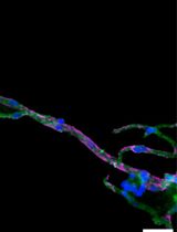
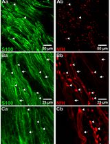
Representative immunofluorescent stained images to show the structures of teased nerve fibers in Figure 3.

Figure 3. Representative immunofluorescent stained images to show the structures of teased nerve fibers. A. A single individual nerve fiber teased from the sciatic nerve of 2-week old rats was double stained with S100 (a specific marker expressed in the cytoplasm of Schwan cells) and neurofilament (NF, a specific marker for showing axon). B. A single individual nerve fiber teased from the sciatic nerve of 4-month old rats was immunostained with S100 to show the detailed structures of Cajal bands (CB) and Schmidt-Lanterman incisures (SLI) which are only exist in adult nerve fibers. PNC, perinuclear cytoplasm; NR, node of Ranvier.
Note: Both of images were stained with DAPI to show the nuclei of Schwann cells.
Recipes
- 10% chloral hydrate
Dissolve 10 g chloral hydrate in 100 ml distilled water and store at 4 °C, protect from light, and use within 1 week after preparation
- 0.9% NaCl
Dissolve 9 g NaCl in 1,000 ml distilled water, store at 4 °C, and use within 1 week after preparation
- 4% PFA
Dilute 40 g PFA in 1,000 ml PBS
Stir at 65 °C until complete dissolution, store at 4 °C, and use within 2 months after preparation
- 0.1% Triton X-100
Dilute 100 μl Triton X-100 into 100 ml PBS, store at RT, and use within 6 months after preparation
- PBST
Dilute 500 μl TWEEN® 20 into 1,000 ml PBS to a solution containing 0.5% TWEEN® 20, store at RT, and use within 2 months after preparation
- Blocking buffer
Dissolve 0.5 g gelatin (5%, w/v) in 10 ml PBS and add 300 μl Triton X-100 (0.3%) to the buffer, store at -20 °C, and use within 6 months after preparation
- 1,000x 4,6-diamidino-2-phenylindole (DAPI)
Dissolve 5 mg DAPI in 5 ml distilled water to make 1,000x stock solution of 1 mg/ml, store at -20 °C, use within 2 years after preparation, and dilute to 1x with PBS before use
Acknowledgments
This protocol is adapted from the previously published paper (Wen et al., 2017). This work was supported by the National Key Basic Research Program of China (2014CB542202 and 2014CB542205), National Natural Science Foundation of China (30973095, 81371354 & 81571182); Science and Technology Project of Guangzhou (12C32121609) and Natural Science Foundation of Guangdong Province (S2013010014697) to J Guo.
References
- Wen, J., Qian, C., Pan, M., Wang, X., Li, Y., Lu, Y., Zhou, Z., Yan, Q., Li, L., Liu, Z., Wu, W. and Guo, J. (2017). Lentivirus-mediated RNA interference targeting RhoA slacks the migration, proliferation, and myelin formation of Schwann cells. Mol Neurobiol 54(2): 1229-1239.
Article Information
Copyright
© 2017 The Authors; exclusive licensee Bio-protocol LLC.
How to cite
Wen, J., Li, L., Tan, D. and Guo, J. (2017). Preparation of Teased Nerve Fibers from Rat Sciatic Nerve. Bio-protocol 7(19): e2572. DOI: 10.21769/BioProtoc.2572.
Category
Neuroscience > Peripheral nervous system > Sciatic nerve
Neuroscience > Cellular mechanisms > Myelin
Cell Biology > Tissue analysis > Tissue isolation
Do you have any questions about this protocol?
Post your question to gather feedback from the community. We will also invite the authors of this article to respond.