- Protocols

- Articles and Issues
- For Authors
- About
- Become a Reviewer

Ustilago maydis Virulence Assays in Maize


Published: Vol 6, Iss 6, Mar 20, 2016 DOI: 10.21769/BioProtoc.1760 Views: 13787
Reviewed by: Marisa RosaPablo Bolanos-VillegasDennis Nürnberg
Original research article
The authors used this protocol in:
Apr 2015
Advertisement


Protocol Collections
Comprehensive collections of detailed, peer-reviewed protocols focusing on specific topics


Related protocols


Abstract
The basidiomycetous smut fungus Ustilago maydis (U. maydis) infects all aerial parts of its host plant maize (Zea mays L.). Infection symptoms are seen in the form of prominent tumors on all aerial parts of maize, after the establishment of a biotrophic interaction with the host usually around 5-6 days post infection (dpi). The fungus colonizes the various developmentally distinct aerial organs at different stages of development to form these prominent symptoms. Although being a biotrophic plant pathogen, U. maydis can easily be cultivated under axenic conditions to produce a standardized inoculum. The infections can be carried out under laboratory conditions by syringe inoculation on all the aerial organs of maize. This protocol has been successfully utilized to infect all the aerial organs of maize and formulate the virulence assays in U. maydis making it an excellent model system to study phyto-pathological investigations (Schilling et al., 2014; Redkar et al., 2015).
Keywords: UstilagoMaterials and Reagents
- 1 ml syringes (Henke Sass-Wolf, catalog number: 5100.200V0 )
- Needles (16 G, 40 mm) (Premier Healthcare & Hygiene, BD microlance, catalog number: 300637 )
- 50 ml Falcon tubes (Sigma-Aldrich, catalog number: T2318 )
- Glass test tubes (Carl Roth GmbH + Co, catalog number: HA79.1 )
- Soil (Frühstorfer Pikiererde T type soil) and Pots
- Ustilago maydis strains
Note: The U. maydis wild-type or any transgenic strains are maintained as glycerol stock in -80 °C for long term storage. Every strain can be retrieved on a PDA plate. - Maize seeds (e.g., variety Early Golden Bantam or Gaspe Flint)
Note: The maize variety Early Golden Bantam is a standard maize line that requires around 4 weeks for the floral switch and development of the male inflorescence. The maize variety Gaspe Flint is a mutant maize that completes its life cycle in 35-38 days and hence has an early floral switch with the development of male inflorescence around 15 days after sowing. Hence this variety is suitable for floral infections with U. maydis. - Yeast extract (BD, Difco, catalog number: 210934 )
- Peptone (BD, Difco, catalog number: 211830 )
- Sucrose (Carl Roth GmbH + Co, catalog number: 9097.2 )
- Double distilled water
- 2.4% Potato Dextrose Broth (BD, Difco, catalog number: 254920 )
- 2% agar (BD, Difco, catalog number: 214010 )
- YEPSL medium (modified from Tsukada et al., 1988) (see Recipes)
- Potato Dextrose Agar (PDA) plates (see Recipes)
Equipment
- Greenhouse
- Erlenmeyer flasks (250 ml)
- Disposable cuvettes (Carl Roth GmbH + Co, catalog number: Y195.1 )
- Incubator [e.g., Certomat BS-1 (B. Braun Biotech International)]
- Centrifuge (Thermo Fisher Scientific)
- Spectrophotometer [e.g. Ultrospec 3000 proUV/Visible (GE Healthcare)]
- Knife
Procedure
- Cultivation of maize plants
- Maize varieties (that may have different lengths of developmental switch) (e.g. cv Early Golden Bantam or cv Gaspe Flint, or your maize cultivar of choice) are grown in round pots of 20 cm with 4 seeds in each pot at equal spacing. The pots are kept in a temperature controlled greenhouse with the following conditions (14-h/10-h light/dark cycle, 28/22 °C. Depending on the growth behavior of the plants, it must be ensured that the right stage is used for infection. Seedling infections are regularly done at the three leaf stage. Tassel infections are done when the tassel floral meristem is in the mitotic phase before the pollen formation (tassel size of 1-1.5 cm).
- Both varieties can be grown in soil in approximately 15 cm round pots with 4 plants each. The variety Gaspe Flint can mainly be used for tassel (male inflorescence) infections as they have an early floral switch (15 days) as compared to the standard maize (early golden Bantam which is approx. 4 weeks).
- Maize varieties (that may have different lengths of developmental switch) (e.g. cv Early Golden Bantam or cv Gaspe Flint, or your maize cultivar of choice) are grown in round pots of 20 cm with 4 seeds in each pot at equal spacing. The pots are kept in a temperature controlled greenhouse with the following conditions (14-h/10-h light/dark cycle, 28/22 °C. Depending on the growth behavior of the plants, it must be ensured that the right stage is used for infection. Seedling infections are regularly done at the three leaf stage. Tassel infections are done when the tassel floral meristem is in the mitotic phase before the pollen formation (tassel size of 1-1.5 cm).
- U. maydis culture preparation
- U. maydis strains growing on a fresh PDA plate (no older than 1 week) are used for preparation of pre-inoculum. The strains are inoculated in a 3 ml YEPSL medium in a sterile glass test tube and incubated overnight at 28 °C with shaking at 200 rpm.
- The pre-inoculum (40 µl) is used for inoculation of the large media (50 ml) in a 250 ml sterile Erlenmeyer flask and then incubated at 28 °C with shaking at 200 rpm for around 10-12 h.
- The cultures are then harvested when the optical density (OD600) reaches to 0.6 to 0.8 (cultures should not be below 0.5, nor above 1.0). This range of OD ensures the growth of the U. maydis sporidia in the active dividing phase. The cultures are then centrifuged in 50 ml Falcon tubes at 900 x g for 5 min at room temperature.
- The harvested cells are resuspended and washed in sterile water to remove all the media remains. The step B4 of centrifugation is repeated.
- The cells are then resuspended by pipetting or vortexing in water to OD600 = 1.0. This inoculum is used to infect the maize plants.
Note: For infection with wild type U. maydis strains (e.g., FB1 and FB2 which are the two compatible mating types) are to be mixed together in equal volume at OD600 =1.0 for a successful infection. In case of the solopathogenic strain SG200 (Kämper et al., 2006) or in case of any mutant resulting from this background one cultural strain can be used as inoculum.
- U. maydis strains growing on a fresh PDA plate (no older than 1 week) are used for preparation of pre-inoculum. The strains are inoculated in a 3 ml YEPSL medium in a sterile glass test tube and incubated overnight at 28 °C with shaking at 200 rpm.
- U. maydis virulence assays
In seedlings:- Seedling infections are done with the maize plantlets that normally show three juvenile leaf stage. For most of the cultivars of maize, the plants are 7 days old when this developmental stage is reached (Figure 1).
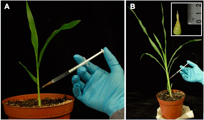
Figure 1. Maize plants cv. Early Golden Bantam and their inoculation with U. maydis. A. Maize seedling at the three leaf stage is inoculated with U. maydis for seedling leave infection. The syringe points into the center of the leaf whirl. B. A maize plant is inoculated with U. maydis for tassel infection. The syringe hits the tassel inside the stem. Inset: the maize tassel at the stage of fungal infection. - The seeding infections are carried out by syringe with 300-500 μl of the inoculum cell suspension into the interior of the leaf whorl. The injection site is chosen approx. 1 cm above the soil, which is about 2.5 to 3 cm above the basal plant meristem and is the juvenile stem.
- The leaf sheaths of the first and second leaf and the leaf blades of the third and fourth leaf which are immersed into the whorl are pierced by the syringe half way onto the center of the stem cylinder, which later show an infection mark after the symptom development.
- Once the inoculum is seen on the inner whorl of leaves the seedling is known to be successfully infected.
- Plants are kept in controlled growth conditions at 28/22 °C. Successful infection will lead to tumor appearance after 4-6 dpi.
- The tumors formed by the artificial inoculation of seedlings are scored at 12 dpi by a modified standard scheme as described previously (Kämper et al., 2006) which is shown in Table 1 and shown below (Figure 2).

Figure 2. Morphological symptoms in seedling (left) and tassel tissues (right) of maize after infection by U. maydis. Photographs were taken at 10 dpi.
Table 1. Classification of symptoms of infected maize seedlingsPlant symptoms Description No symptom The plant shows no signs of infection Chlorosis The plant shows chlorotic discolouration of the infected leaves (third leaf and younger) Small tumors The largest tumors of the plant are <1.5 mm Normal tumors Tumors of the plant are 2-4 mm in diameter Heavy Tumors Very strong tumors leading to a stunted growth of stem Dead Plant The plant is dead and looks necrotic after infection with U. maydis
- Tassel infections are done immediately after floral switch of the plant when the tassel is still in the actively dividing mitotic phase. In cv. Gaspe Flint this is normally after 15 days and after around 4 weeks in cv. Early Golden Bantam after sowing (see Figure 1).
- The tassel infections are also performed using a syringe based method. The immature tassels location is identified without cutting the plant by pressing the stem. The tassel meristem embedded in the stem has an air space surrounding it which makes the stem hollower. Above the tassel which terminates the shoot, the leaf sheaths overlap, but there is no solid center and this region contains the developing inflorescence.
- Prior to injection, one or two plants are cut open with a sharp knife from the base, to understand the position of the tassel meristem that is embedded in the whorl of leaves.
- Based on this height, the tassel location is approximately identified on all plants. The standard injection protocol involves inoculation of 1 ml of culture injected all at once, taking care to penetrate only halfway across the plant diameter. This ensures the inoculum to the entire floral meristem and fills the air space around the tassel.
- Plants are kept in a controlled greenhouse at 28 °C which show successful tumor appearance after 8-10 dpi (Figure 2).
- Disease symptoms in the tassels are scored 10 days after infection in cv. Gaspe Flint and after 14 days in cv. Early Golden Bantam by the scoring scheme described previously (Schilling et al., 2014, Table 2) and shown below.
Table 2. Classification of symptoms of infected maize tasselsTassel Symptom Description Tumors<50% of tassel, small The part of the tassel is converted into small tumors in range of 2-4 mm Tumors<50% of tassel, large Less than half of the tassel is converted into large tumors >4 mm Tumors>50% of tassel, small The entire tassel inflorescence is covered with small tumors. Tumors>50% os tassel, large The complete tassel is converted into heavy tumors by U. maydis Tassel growth stunted The tassel is arrested at the developmental time point at which it was infected and this 1-3 cm stunted tassel becomes tumorous.
- Seedling infections are done with the maize plantlets that normally show three juvenile leaf stage. For most of the cultivars of maize, the plants are 7 days old when this developmental stage is reached (Figure 1).
Recipes
- YEPSL medium
This medium consists of 0.4% (w/v) yeast extract, 0.4% (w/v) peptone, 2% (w/v) sucrose in double distilled water. - PDA plates
The PDA medium consists of 2.4% Potato Dextrose Broth and 2% agar in double distilled water. After autoclaving approx. 18 ml of media in each petri plate.
Acknowledgments
Our work was funded by the Max Planck Society, The Deutsche Forschungsgemeinschaft (DFG), The Deutscher Akademischer Austauschdienst (DAAD) and the Cluster of Excellence on Plant Science (CEPLAS). The protocol is adapted from Kämper et al. (2006); Schilling et al. (2014) and Redkar et al. (2015).
References
- Kamper, J., Kahmann, R., Bolker, M., Ma, L. J., Brefort, T., Saville, B. J., Banuett, F., Kronstad, J. W., Gold, S. E., Muller, O., Perlin, M. H., Wosten, H. A., de Vries, R., Ruiz-Herrera, J., Reynaga-Pena, C. G., Snetselaar, K., McCann, M., Perez-Martin, J., Feldbrugge, M., Basse, C. W., Steinberg, G., Ibeas, J. I., Holloman, W., Guzman, P., Farman, M., Stajich, J. E., Sentandreu, R., Gonzalez-Prieto, J. M., Kennell, J. C., Molina, L., Schirawski, J., Mendoza-Mendoza, A., Greilinger, D., Munch, K., Rossel, N., Scherer, M., Vranes, M., Ladendorf, O., Vincon, V., Fuchs, U., Sandrock, B., Meng, S., Ho, E. C., Cahill, M. J., Boyce, K. J., Klose, J., Klosterman, S. J., Deelstra, H. J., Ortiz-Castellanos, L., Li, W., Sanchez-Alonso, P., Schreier, P. H., Hauser-Hahn, I., Vaupel, M., Koopmann, E., Friedrich, G., Voss, H., Schluter, T., Margolis, J., Platt, D., Swimmer, C., Gnirke, A., Chen, F., Vysotskaia, V., Mannhaupt, G., Guldener, U., Munsterkotter, M., Haase, D., Oesterheld, M., Mewes, H. W., Mauceli, E. W., DeCaprio, D., Wade, C. M., Butler, J., Young, S., Jaffe, D. B., Calvo, S., Nusbaum, C., Galagan, J. and Birren, B. W. (2006). Insights from the genome of the biotrophic fungal plant pathogen Ustilago maydis. Nature 444(7115): 97-101.
- Redkar, A., Hoser, R., Schilling, L., Zechmann, B., Krzymowska, M., Walbot, V. and Doehlemann, G. (2015). A secreted effector protein of Ustilago maydis guides maize leaf cells to form tumors. Plant Cell 27(4): 1332-1351.
- Schilling, L., Matei, A., Redkar, A., Walbot, V. and Doehlemann, G. (2014). Virulence of the maize smut Ustilago maydis is shaped by organ-specific effectors. Mol Plant Pathol 15(8): 780-789.
- Tsukada, T., Carleton, S., Fotheringham, S. and Holloman, W. K. (1988). Isolation and characterisation of an autonomously replicating sequence from Ustilago maydis. Mol Cell Biol 8: 3703-3709.
Article Information
Copyright
© 2016 The Authors; exclusive licensee Bio-protocol LLC.
How to cite
Redkar, A. and Doehlemann, G. (2016). Ustilago maydis Virulence Assays in Maize. Bio-protocol 6(6): e1760. DOI: 10.21769/BioProtoc.1760.
Category
Plant Science > Plant immunity > Disease bioassay
Microbiology > Microbe-host interactions > Fungus
Microbiology > Microbe-host interactions > In vivo model > Plant
Do you have any questions about this protocol?
Post your question to gather feedback from the community. We will also invite the authors of this article to respond.