- Protocols

- Articles and Issues
- For Authors
- About
- Become a Reviewer

Fluoro-Jade B Staining for Neuronal Cell Death

Published: Vol 6, Iss 1, Jan 5, 2016 DOI: 10.21769/BioProtoc.1702 Views: 24002
Reviewed by: Jia LiSabine Le SauxAnonymous reviewer(s)
Original research article
The authors used this protocol in:
Apr 2015

Protocol Collections
Comprehensive collections of detailed, peer-reviewed protocols focusing on specific topics


Related protocols


Abstract
Fluoro-Jade is a fluorescent derivative used for histological staining of degenerating neurons. This technique is simple and sensitive enough to label distal dendrites, axons, axon terminals as well as neuronal bodies. Fluoro-Jade has excitation and emission peak of 480 and 525 nanometer respectively. It can be visualized using a fluorescein/FITC filter. Some reports have demonstrated that Fluoro-Jade can also be useful to detect glial cell death (Anderson et al., 2013; Damjanac et al., 2007).
Keywords: Fluoro-Jade BMaterials and Reagents
- Superfrost plus Microscope slide (Thermo Fisher Scientific, catalog number: 12-550-17 )
- Cover Glass (Thermo Fisher Scientific, catalog number: 12-545-88 )
- Tissue sample
- Fluoro-Jade B (Merck Millipore Corporation, catalog number: AG310 )
- Paraformaldehyde (Electron Microscopy Science, catalog number: 19210 )
- Potassium permanganate (KMnO4) (Sigma-Aldrich, catalog number: 223468 )
- DAPI (Life Technologies, catalog number: D3571 )
Note: Currently, it is “Thermo Fisher Scientific, Molecular ProbesTM, catalog number: D3571”. - Glacial Acetic acid (CH3CO2H) (Sigma-Aldrich, catalog number: A9967 )
- Ethanol
- Xylene (Sigma-Aldrich, catalog number: 534056 )
- Sodium hydroxide (NaOH) (Sigma-Aldrich, catalog number: S8045 )
- Sodium tetraborate decahydrate (Sigma-Aldrich, catalogue number: B9876 )
- Sodium chloride (NaCl) (Sigma-Aldrich, catalog number: S3014 )
- potassium phosphate dibasic (K2HPO4) (Sigma-Aldrich, catalog number: P3786 )
- potassium phosphate monobasic (KH2PO4) (Sigma-Aldrich, catalog number: P9791 )
- 4% paraformaldehyde (see Recipes)
- KPBS (see Recipes)
- 0.2% Fluoro-Jade (see Recipes)
- Fluoro-Jade solution (see Recipes)
- 0.2% DAPI (see Recipes)
Equipment
- Vacuum Desiccators (Thermo Fisher Scientific, catalog number: 08-642-5 )
- Tissue-Tek slide staining set (Electron Microscopy Science, catalog number: 62540-01 )
- 24 slide holder (Electron Microscopy Science, catalog number: 62543-06 )
- Orbital shaker
- Timer
- Slide Warmer
- DPX mounting medium (a mixture of the polystyrene distyrene and the plasticizer dibutylphthalate) (Electron Microscopy Science, catalog number: 13512 )
Procedure
- Mount tissue sections (20-35 μm cut on microtome or cryostat) on Superfrost plus slide and let the slide dry overnight under vacuum.
- Fix the tissue on slide warmer 30 min at 60 °C and/or 20 min in paraformaldehyde 4%.
- Follow by 2 min in KPBS.
Note: All the following steps are done at room temperature. - Dehydrate in 50%-70%-100% Ethanol 2 min each.
- Rehydrate by going back in 70%-50% Ethanol and KPBS, 2 min each.
- Incubate in potassium permanganate 0.06% (dilute in water) 5 min at room temperature.
- Rinse in water 1 min.
- Incubate in Fluoro-Jade solution at room temperature for 10 min and gently shake on orbital shaker or by doing several dips during incubation (three dips of few seconds 3 times during incubation). Use opaque cup for the Fluoro-Jade incubation and keep the slide in shelters from light for the rest of the procedure.
Note: Fluoro-Jade solution and potassium permanganate 0.06% must be prepared fresh. - Follow by three rinses of 1 min each in water.
- Dry the slides overnight under vacuum at room temperature.
- Clear the slide in Xylene (3 x 2 min).
- Cover slip with DPX and dry 24-48 h under hood before microscope analysis.
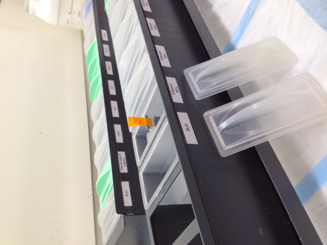
Figure 1. Example of the setup use for incubation and/or dipping of the slides in different solutions
Representative data

Figure 2. FJB-positive neurons in the mouse cerebral cortex following ischemic stroke
Notes
To ensure reproducibility between protocols, use the same method of tissue preparation.
Recipes
- 4% paraformaldehyde
Heat 700 ml distilled water at 65 °C
Add 40 g paraformaldehyde
5 ml NaOH
When Paraformaldehyde is completely dissolved add 38 g sodium tetraborate
Complete at 1 liter with distilled water - KPBS
3.81 g Potassium phosphate dibasic
0.45 g Potassium phosphate monobasic
8.1 g sodium chloride
Complete to 1 liter with distilled water - 0.2% Fluoro-Jade
Dilute 50 mg Fluoro-Jade B in 25 ml of sterile water and aliquot
Keep this stock solution at -20 °C in shelters from light - Fluoro-Jade solution
Fluoro-Jade B 0.0004%
Acetic acid 0.1%
DAPI 0.0001% in water - 0.2% DAPI
Dilute 10 mg DAPI in 5 ml of sterile water and aliquot
Keep this stock solution at 4 °C in shelters from light
Acknowledgments
This protocol was adapted from Schmued et al. (1997). This work was supported by grants from the Canadian Institutes for Health Research (CIHR) and the Multiple Sclerosis Scientific Research Foundation of Canada.
References
- Anderson, K. J., Fugaccia, I. and Scheff, S. W. (2003). Fluoro-Jade B stains quiescent and reactive astrocytes in the rodent spinal cord. J Neurotrauma 20(11): 1223-1231.
- Damjanac, M., Rioux Bilan, A., Barrier, L., Pontcharraud, R., Anne, C., Hugon, J. and Page, G. (2007). Fluoro-Jade B staining as useful tool to identify activated microglia and astrocytes in a mouse transgenic model of Alzheimer's disease. Brain Res 1128(1): 40-49.
- Schmued, L. C., Albertson, C. and Slikker, W., Jr. (1997). Fluoro-Jade: a novel fluorochrome for the sensitive and reliable histochemical localization of neuronal degeneration. Brain Res 751(1): 37-46.
Article Information
Copyright
© 2016 The Authors; exclusive licensee Bio-protocol LLC.
How to cite
Laflamme, N., Préfontaine, P. and Rivest, S. (2016). Fluoro-Jade B Staining for Neuronal Cell Death. Bio-protocol 6(1): e1702. DOI: 10.21769/BioProtoc.1702.
Category
Neuroscience > Development > Histological staining
Cell Biology > Cell staining > Whole cell
Do you have any questions about this protocol?
Post your question to gather feedback from the community. We will also invite the authors of this article to respond.