- Protocols

- Articles and Issues
- For Authors
- About
- Become a Reviewer

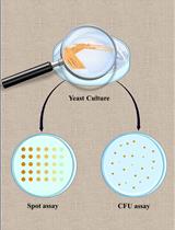
Spot Assay for Yeast

Published: Vol 2, Iss 1, Jan 5, 2012 DOI: 10.21769/BioProtoc.16 Views: 42813
Advertisement


Protocol Collections
Comprehensive collections of detailed, peer-reviewed protocols focusing on specific topics


Abstract
This protocol can be used to compare the cell growth rate of yeast under different growth conditions. It involves the serial dilution and spotting of yeast colonies.
Materials and Reagents
- Yeast cells
- YES medium
Equipment
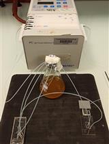
- Multichannel Pipetman (Eppendorf)
- OmniTray (V&P Scientific)
- Microfuge tube
- Standard laboratory spectrophotometer
Procedure
- Start cultures from a 2 day old plate. Use pipette tip to pick up strains and resuspend them in 1.5 ml YES medium or water.
- Vortex and transfer 1 ml to another microfuge tube. Test OD600 for an accurate reading, the OD should be between 0.1 and 0.5.
- Dilute the rest of the suspension to 16 OD600, around 1.6 x 10⁶ cells per ml, 1,600 cells per μl. That is 4,800 or 5,000 cells per 3 μl. Spot 3 μl cells on each position.
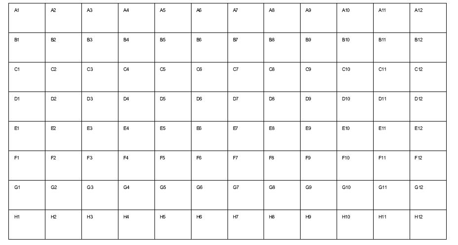
- If using OmniTray, start with the 1st column (8 wells in each column).
- Do 5 fold serial dilution from the 1st to 5th column. Leave the 6th column empty. Transfer another 8 strains culture into the 7th column and do another 5 fold dilution.
Acknowledgments
This protocol has been modified and adapted in the Espenshade Lab, Johns Hopkins School of Medicine. Funding to support different projects that have used this protocol has come from NIH – National Heart, Lung, and Blood Institute, National Institute of Allergy and Infectious Diseases, the Pancreatic Cancer Action Network, and the American Heart Association.
References
- Tong, Z., Gao, X. D., Howell, A. S., Bose, I., Lew, D. J. and Bi, E. (2007). Adjacent positioning of cellular structures enabled by a Cdc42 GTPase-activating protein-mediated zone of inhibition. J Cell Biol 179(7): 1375-1384.
Article Information
Copyright
© 2012 The Authors; exclusive licensee Bio-protocol LLC.
How to cite
Editor, B. (2012). Spot Assay for Yeast. Bio-protocol 2(1): e16. DOI: 10.21769/BioProtoc.16.
Category
Microbiology > Microbial cell biology > Cell viability
Microbiology > Microbial cell biology > Cell isolation and culture
Cell Biology > Cell isolation and culture > Cell growth
Do you have any questions about this protocol?
Post your question to gather feedback from the community. We will also invite the authors of this article to respond.