- Protocols

- Articles and Issues
- For Authors
- About
- Become a Reviewer

Murine in vivo CD8+ T Cell Killing Assay
Published: Vol 4, Iss 13, Jul 5, 2014 DOI: 10.21769/BioProtoc.1172 Views: 19658
Reviewed by: Omar AkilAnonymous reviewer(s)
Original research article
The authors used this protocol in:
Aug 2013
Advertisement


Protocol Collections
Comprehensive collections of detailed, peer-reviewed protocols focusing on specific topics


Related protocols
Abstract
Antigen-specific killing ability of effector CD8+ T cells is critical for protective immunity against infection. Here, we describe in vivo cytotoxic T cell assay to examine effector function of antigen-specific CD8+ T cells. Mice infected with Listeria monocytogenes (L. monocytogenes) expressing chicken ovalbumin as a model antigen mount ovalbumin-specific CD8+ T cell responses. Effector CD8+ T cell function in vivo is determined by mixed transfer of OVA peptide-pulsed target cells with control target cells into the previously immunized mice. Difference in CFSE expression levels clearly marks two distinct populations: Antigen-pulsed target cells-CFSElow vs. unpulsed target cells-CFSEhi. The frequencies between antigen-pulsed target cells and control target cells are used as readouts of antigen-specific killing.
Materials and Reagents
- Splenocytes from a wild type mouse
- PBS (Thermo Fisher Scientific, catalog number: BP399-20 )
Note: 10x solution, diluted to 1x in house in distilled water and sterilized by autoclave.
- RBC lysis buffer (eBioscience, catalog number: 00-4333-57 )
- HBSS without Ca2+ and Mg2+ (Life Technologies, Gibco®, catalog number: 14175-095 )
- RPMI-1640 medium (Life Technologies, Gibco®, catalog number: 11875-119 )
- Fetal bovine Serum (Atlanta Biologicals, catalog number: S11055H )
- Penicillin/streptomycin (Gemini Bio-Products, catalog number: F52M00E )
- L-Glutamine (Life Technologies, Gibco®, catalog number: 25030-081 )
- Trypan blue solution (Life Technologies, Gibco®, catalog number: 15250-061 )
- OVA257-264 synthetic peptide (Sigma-Aldrich, catalog number: S7951 )
- 5(6)-Carboxyfluorescein diacetate N-succinimidyl ester (CFSE) (Sigma-Aldrich, catalog number: 21888 )
- Dimethyl sulfoxide (DMSO) (Sigma-Aldrich, catalog number: D-8418 )
- Collagenase D (Sigma-Aldrich, catalog number: C-5138 )
- Percoll (Sigma-Aldrich, catalog number: P-1644 )
- Complete RPMI-1640 media (see Recipes)
- 100% percoll solution (see Recipes)
Equipment
- Centrifuge (Thermo Fischer Scientific, SorvallTM Legend RT )
- 37 °C water bath
- Hemocytometer
- 15 ml and 50 ml Falcon tubes
- 6 well plates (USA Scientific, CytoOne®, catalog number: CC7682-7506 )
- BD LSRII Flow Cytometer (BD)
- 70 µm cell strainer (BD Biosciences, Falcon®, catalog number: 352350 )
- 5 ml polystyrene round-bottom tubes with cell-strainer cap (BD Biosciences, Falcon®, catalog number: 352235 )
- 3 ml syringe (BD, catalog number: 14-823-435 )
Procedure
- Target cell preparation under sterile tissue culture conditions
This step is for preparing peptide-pulsed target cells and stain cells with CFSE to distinguish peptide-pulsed target cells from control target cells.- OVA257-264 peptide-loading for the target cells.
- Splenocytes are RBC lysed followed by washing with PBS twice.
- Resuspend cells in RPMI-1640 complete medium.
- Count the mononuclear cells by trypan blue exclusion using a hemocytometer.
- Resuspend cells at 5 x 10⁶/ml of RPMI-1640 complete medium.
- Divide the cells equally into two separate 50 ml Falcon tubes- one for peptide-pulsed target cells, and the other for unpulsed target cells.
- Add OVA257-264 peptide at 1 µl/ml from a 200 µM stock to peptide-pulsed target cells.
- Add an equivalent amount of PBS to the unpulsed target cells.
- Incubate the cells in a 37 °C water bath for 1 h.
- Wash cells twice with RPMI-1640 complete medium.
- Centrifuge the cells at 1,500 rpm for 3 min at 4 °C.
- Resuspend the cell pellet in HBSS.
- Splenocytes are RBC lysed followed by washing with PBS twice.
- CFSE cell labeling under sterile tissue culture conditions.
- Count all live cells by trypan blue exclusion using hemocytometer.
- Resuspend the cells in HBSS at 5 x 10⁷/ml.
- Thaw an aliquot of 5 mM stock CFSE solution.
- Make a fresh CFSElow stock solution by diluting 5 mM stock 1:10 in DMSO (a final concentration of 0.5 mM).
- Incubate the unpulsed target splenocytes with the higher concentration of CFSE (CFSEhigh): Add 1 µl of the 5 mM stock CFSE for each milliliter of unpulsed target cells (final concentration of 5 µM).
- Incubate the pulsed target splenocytes with the lower concentration of CFSE (CFSElow): Add 1 µl of the 0.5 mM stock CFSE for each milliliter of peptide-pulsed cells (final concentration of 0.5 µM).
- Pipette cells up and down to mix well and incubate in water bath for 10 min at 37 °C. Gently agitate the cells periodically.
- Add 10x the volume of pre-warmed RPMI-1640 complete medium to the CFSE-labeled cells to stop the reaction.
- Pellet cells at 1,500 rpm for 3 min at 4 °C.
- Remove the supernatant and resuspend the pellet in cold RPMI-1640 complete medium.
- Wash the cells two more times with cold RPMI-1640 complete medium.
- Count the mononuclear cells by trypan blue exclusion using a hemacytometer.
- Wash the cells with cold PBS.
- Resuspend each cell populations in PBS at 6.7 x 10⁶/ml.
- Combine an equal volume (~equal numbers) of peptide-pulsed CFSElow cells with unpulsed CFSEhi cells and proceed with flow cytometry analysis (Figure 1a).
- Count all live cells by trypan blue exclusion using hemocytometer.
- OVA257-264 peptide-loading for the target cells.
- Intravenous injection of target splenocytes
To investigate OVA257-264-specific CD8+ T cell killing ability, peptide-pulsed and unpulsed target cells were mixed at a 1:1 ratio and transferred to the previously immunized mice.- Recipient mice were infected with 5,000 colony-forming units (CFU) of Listeria monocytogenes expressing chicken ovalubmin (LM-OVA) 7 days intravenously before the CFSE-labeled cell injection.
- Inject intravenously 300 µl of the combined cell populations into the tail vein of each recipient. Each recipient should receive approximately 1 x 10⁷ peptide-pulsed target cells combined with 1 x 10⁷ unpulsed target cells.
- Wait for 4 h.
- Recipient mice were infected with 5,000 colony-forming units (CFU) of Listeria monocytogenes expressing chicken ovalubmin (LM-OVA) 7 days intravenously before the CFSE-labeled cell injection.
- Preparation of splenocytes and lymphocytes in the liver for flow cytometry analysis
This step is analyzing antigen-specific killing ability in the liver and the spleen by flow cytometric analysis of CFSElow and CFSEhi cell populations.- Prepare splenocytes for flow cytometry analysis.
- Wash once with PBS, and 2-300 µl into a 5 ml round bottom tubes through the cell strainer.
- Isolate lymphocytes from the liver.
- Harvest livers from the recipients and place them on ice.
- Make a fresh collagenase D solution by diluting 20 mg/ml stock 1:20 in PBS (a final concentration of 1 mg/ml).
- Chop liver with a blade on a slide glass and transfer them into a 50 ml falcon tube.
- Add 7 ml of collagenase D (1 mg/ml) and vortex well.
- Incubate in water bath for 30 min at 37 °C. Vortex every 15 min.
- Put tubes on ice and add supernatant on 70 µm filter on a well of a 6 well plate. Grind chunks of the chopped liver with flat portion of 3 ml syringe. Wash the tube with 5 ml of PBS and repeat grinding.
- Transfer them to a 50 ml Falcon tube and spin down at 2,000 rpm for 5 min at 4 °C.
- Make fresh 44% and 66% percoll solution: Make 44% final concentration by diluting 100% percoll in PBS, and make 66% final concentration by diluting 100% percoll in RPMI-1640 medium.
- Resuspend pellet in 7 ml of 44% percoll, and load them on 3 ml of 66% percoll in 15 ml tube.
Note: The gradient separation is sensitive to agitation. Try not to shake the tube.
- Centrifuge at 3,000 rpm for 30 min at 4 °C without brake.
- Transfer the interphase lymphocytes to a new 15 ml tube.
- Pellet cells at 2,000 rpm for 5 min at 4 °C.
- Wash once with PBS, and transfer 2-300 µl into a 5 ml round bottom tubes through the cell strainer.
- Harvest livers from the recipients and place them on ice.
- Proceed to flow cytometry analysis with spleen and liver samples (Figure 1b).
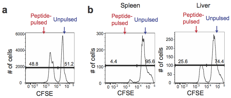
Figure 1. CFSE expression in antigen-pulsed target cells and unpulsed target cells. a. Peptide-pulsed CFSElow and unpulsed CFSEhi splenocytes were mixed at a 1:1 ratio before transferring to the recipients. b. The mixture of peptide-pulsed and unpulsed splenocytes was transferred into the mice predisposed with LM-OVA at day 7 post infection. The spleen and the liver of the recipients were harvested after 4 h to determine percentages of CFSElow and CFSEhi cells among CFSE+ cells.
- Prepare splenocytes for flow cytometry analysis.
Recipes
- Complete RPMI-1640 medium
10% FBS
1% Penicillin/streptomycin with L-Glutamine
- 100% percoll solution
90% of percoll
10% of 10x PBS
Notes
- The gradient separation is sensitive to agitation. Try not to shake the tube.
Acknowledgments
The protocol was adapted from a previously described study (Manjunath et al., 2001). This work was supported by the Starr Cancer Consortium (13-A123 to M.O.L. and M.Q.Z.), the Rita Allen Foundation (M.O.L.), the NBRPC (2012CB316503 to M.Q.Z), and the NIH (HG001696 to M.Q.Z.).
References
- Ingulli, E. (2007). Tracing tolerance and immunity in vivo by CFSE-labeling of administered cells. Methods Mol Biol 380: 365-376.
- Kim, M. V., Ouyang, W., Liao, W., Zhang, M. Q. and Li, M. O. (2013). The transcription factor Foxo1 controls central-memory CD8+ T cell responses to infection. Immunity 39(2): 286-297.
Article Information
Copyright
© 2014 The Authors; exclusive licensee Bio-protocol LLC.
How to cite
Kim, M. V., Ouyang, W., Liao, W., Zhang, M. Q. and Li, M. O. (2014). Murine in vivo CD8+ T Cell Killing Assay. Bio-protocol 4(13): e1172. DOI: 10.21769/BioProtoc.1172.
Category
Immunology > Immune cell function > Antigen-specific response > Membrane protein detection
Immunology > Animal model > Mouse
Microbiology > Microbe-host interactions > In vivo model > Mammal
Do you have any questions about this protocol?
Post your question to gather feedback from the community. We will also invite the authors of this article to respond.