


- Protocols

- Articles and Issues
- For Authors
- About
- Become a Reviewer


Separating Chromera velia Zoospores From Culture and Estimating Their Average Motility Speed and Lifespan



Published: Vol 16, Iss 14, Jul 20, 2026 DOI: 10.21769/BioProtoc.5756 Views: 213
Reviewed by: Veronika KselíkováAnonymous reviewer(s)
 PDF
PDF  Ask a question
Original research article
The authors used this protocol in:

Dec 2023

Protocol Collections
Comprehensive collections of detailed, peer-reviewed protocols focusing on specific topics




Related protocols
Abstract
Chromera velia is an apicomplexan alga uniquely positioned as the closest photosynthetic relative to apicomplexan parasites (Sporozoa), which include the human pathogens that cause malaria (Plasmodium) and toxoplasmosis (Toxoplasma). Under favorable conditions, C. velia forms motile zoospores that contribute to dispersal and possibly host interaction. However, zoospores coexist with other developmental stages in culture, making their isolation technically challenging. Previous studies characterized the phototactic behavior of zoospores in several taxa, yet this response has not been used to separate motile zoospores from mixed cultures. Other reported methods for zoospore recovery relied instead on physical or chemical principles such as passive filtration, differential centrifugation, or column-based purification, all of which can compromise zoospore motility and viability through mechanical shear or osmotic changes. To address this limitation, we developed a non-invasive, simple, and effective method for rapid zoospore isolation depending entirely on their negative phototaxis response. Using a directional light gradient, the method enables reliable collection of active, motile zoospores without specialized equipment or chemical treatments. Our protocol is straightforward to reproduce, relies on standard laboratory equipment, can be completed in under two hours, and yields a zoospore fraction of sufficient quality for live-imaging, motility assays, and downstream molecular and -omics applications. It may also be adapted to other flagellated protists with light-responsive motile stages.
Key features
• Requires basic experience with algal culturing and access to a light-controlled environment.
• A typical 7-day pre-cultivation period is needed for Chromera velia to initiate and reach peak zoosporogenesis.
• Provides a rapid procedure for isolating C. velia zoospores that can be fully completed within 1–2 h, including material preparation.
• Enables downstream experiments with clean, motile zoospore populations for physiological, behavioral, molecular, or -omics analyses.
Keywords: Chromera velia Zoospore isolation
Negative phototaxis
Motility assay
Life cycle
Zoospore isolation
Negative phototaxis
Motility assay
Life cycle
Graphical overview
Background
Sitting at the evolutionary crossroads between photosynthesis and parasitism, Chromera velia (Apicomplexa, Apicomonada [1]) is a photoparasitic alga and the closest known photosynthetic relative of apicomplexan parasites, the group encompassing the causative agents of malaria (Plasmodium) and toxoplasmosis (Toxoplasma). Chromera velia has been isolated from the scleractinian coral Plesiastrea purpurea using a method developed for isolating intracellular coral symbionts. These symbionts, usually dinoflagellates, reside within coral tissue and support the host by supplying photosynthetically derived carbon, receiving carbon dioxide and protection in return. Chromera velia was initially presumed to have a similar life strategy [2]. However, Cumbo et al. [3] demonstrated that, unlike dinoflagellate symbionts that invade the endoderm of coral larvae via the oral pore, C. velia was also found in the ectoderm, indicating active penetration from the outside. Additionally, transcriptomic analyses of C. velia–infected coral larvae revealed expression patterns indicative of a host–pathogen interaction rather than a mutualistic association [4].
Chromera velia has a complex life cycle that is fully maintained under laboratory conditions (Figure 1). It includes immotile stages such as coccoid cells (Figure 2A), autosporangia, zoosporangia (Figure 2B), and cysts, as well as highly motile, biflagellate zoospores (Figure 2C). The process that produces zoospores is called zoosporogenesis [5]. The zoospore is the only life stage believed to penetrate host cells, as it contains a primitive apical complex, in the form of a conoid (pseudoconoid), a structure known from parasitic apicomplexans [6,7]. Thus, C. velia plays a significant role in our understanding of the evolutionary origin of parasitism [8].
Beyond C. velia, zoosporogenesis and the release of motile cells depend heavily on the biology and life cycle of each organism and its adaptation to its environment. The release of zoospores can be triggered by various biotic and abiotic factors, including osmotic stress, salinity, temperature, pH, nutrient availability, wave motion, and light intensity [9]. In C. velia, zoosporogenesis is induced particularly in response to specific light wavelengths and medium composition [5,10]. Appearance of zoospores has been well documented under 12/12 h light/dark (LD) conditions [2,5]. Zoospore numbers peak between 7 and 10 days after culture inoculation and 6 h after the onset of illumination [6,10]. Except under red light, C. velia zoospores display negative phototaxis in response to all tested light spectra, actively swimming away from the light source. Their average swimming speed is estimated at approximately 2 cm/min [10]. A persistent challenge in the study of C. velia has been the reliable isolation of clean zoospore populations, free from coccoid and other non-motile stages, which is crucial for downstream applications such as gene expression analysis, behavioral assays, and physiological studies. The phototactic behavior of zoospores has been characterized in several taxa, including Ulkenia sp. zoospores [11], Allomyces sp. [12], and Ulva sp. [13]; however, these studies focused on identifying the wavelengths eliciting positive or negative phototaxis and did not exploit this response as a separation principle. To our knowledge, the present protocol is the first to use phototaxis directly as a means of purifying motile zoospores from a mixed culture. Previously reported methods for recovering zoospores or other flagellated stages in related systems rely instead on physical or chemical principles. Passive filtration through nylon or polycarbonate meshes has been used to collect zoospores in oomycetes and chytrids [14,15], differential centrifugation has been applied to actinomycete zoospores [16], and column-based purification, such as anion-exchange chromatography of Eimeria sporozoites [17], has been conducted, all of which can compromise zoospore motility and viability by mechanical shear or osmotic change. By contrast, our method is non-invasive, and its selectivity is driven entirely by the behavior of the zoospores—negative phototaxis. Using a directional light source and a standard U-shaped glass tube, only actively swimming zoospores migrate into the collection arm of the U-tube, yielding a population of intact, motile cells suitable for live-cell imaging, motility assays, and downstream molecular analyses. Furthermore, this low-cost approach may be adapted for other flagellated protists exhibiting light-responsive motility.
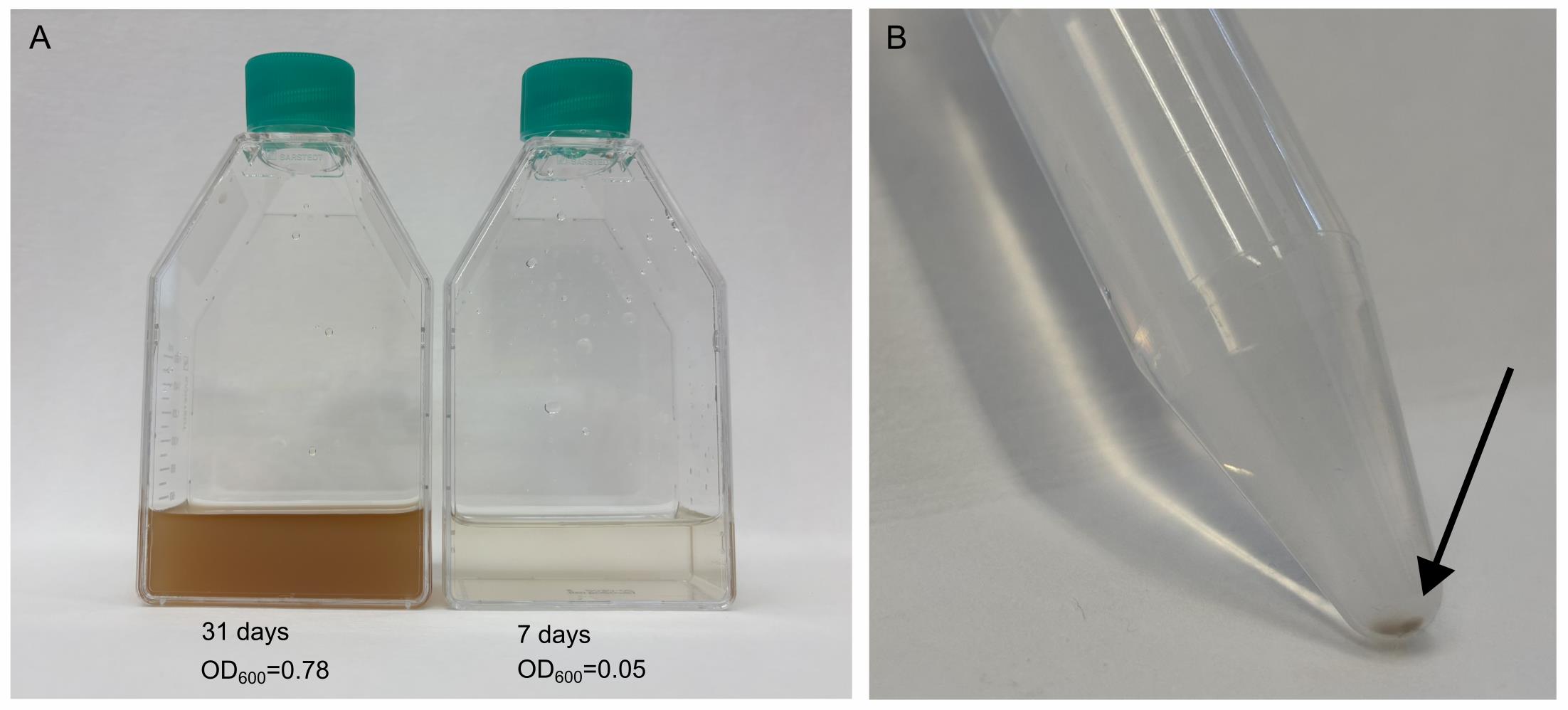
Figure 1. Representative stages of Chromera velia cultures and isolated zoospores. (A) A 31-day-old stationary culture (OD600 = 0.78) was used as inoculum, compared with a 7-day-old early exponential culture (OD600 = 0.05) enriched in zoospores used for isolation. (B) Concentrated zoospore fraction visible as a brownish layer at the tip of the tube after isolation (arrow).
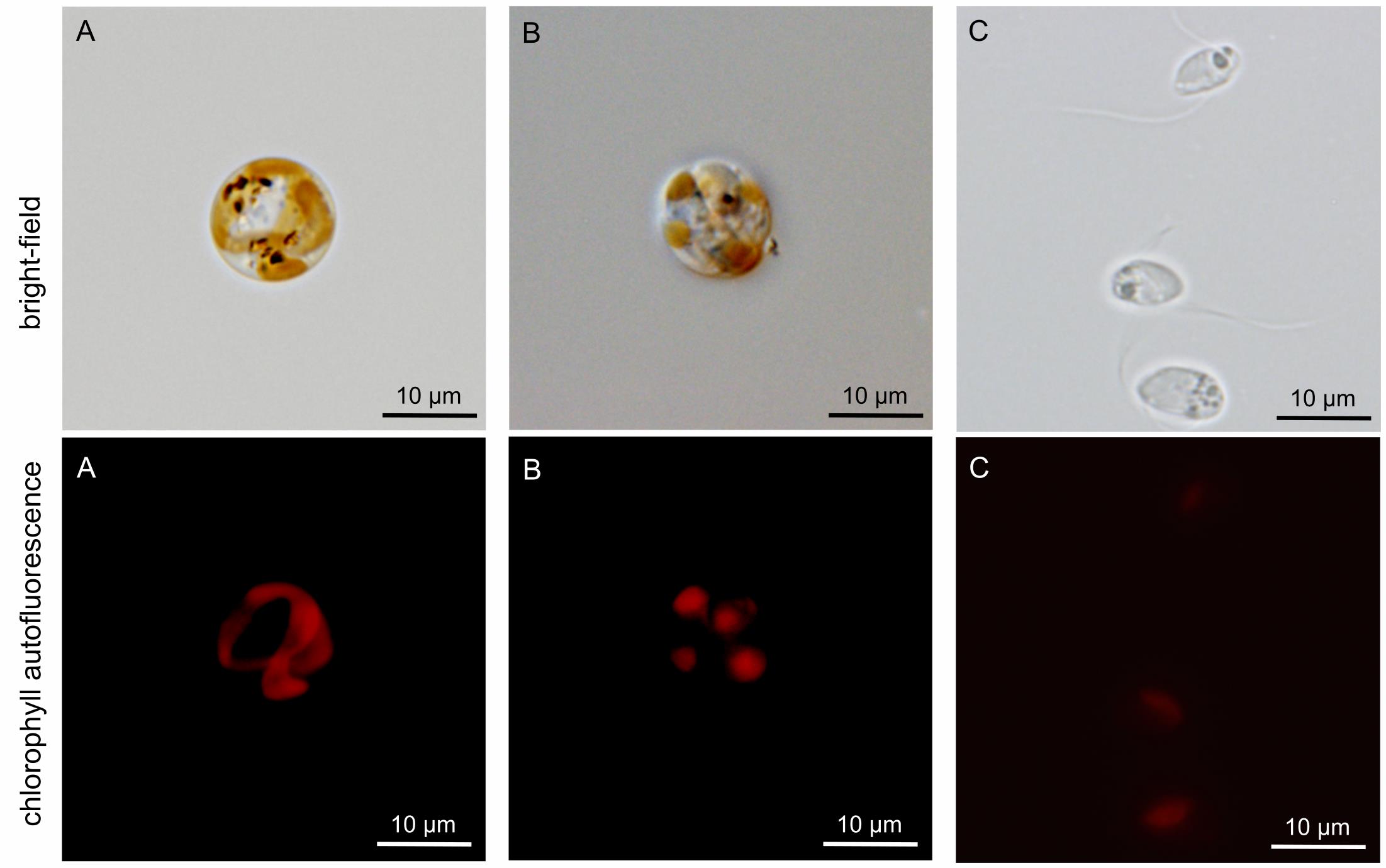
Figure 2. Brightfield light microscopy and chlorophyll autofluorescence of Chromera velia developmental stages. Upper row: Brightfield images. Lower row: Corresponding chlorophyll autofluorescence (red signal). (A) Coccoid cell containing a single, well-developed plastid. (B) C. velia zoosporangium containing six zoospores. (C) Isolated zoospores fixed with 4% paraformaldehyde.
Materials and reagents
Biological materials
1. Chromera velia [2] (National Centre for Marine Algae and Microbiota, Maine, USA; catalog number: CCMP 2878)
Reagents
1. Sea salt: Tropic Marin PRO REEF (Tropic Marin, catalog number: 10581)
2. Guillard F/2 marine enrichment solution (Sigma-Aldrich, catalog number: G0154)
3. Distilled water (Milli-Q or equivalent)
4. Azoxystrobin (Azo) (TraceCERT®) (Sigma-Aldrich, catalog number: 43854)
5. Dimethyl sulfoxide (DMSO) (≥99.5%, suitable for plant cell culture) (Sigma-Aldrich, catalog number: D4540)
6. Ethanol 70% (v/v), prepared from absolute or 96% ethanol with distilled water; for surface sterilization, typical exposure time 1–2 min (optional)
7. Bleach solution 1% (v/v), prepared by diluting commercial bleach stock solution [Savo Original, Unilever; ~3%–5% (w/v) sodium hypochlorite] 1:5 in distilled water (optional)
Solutions
1. Guillard’s medium (F/2 medium) (see Recipes)
2. Azoxystrobin stock solution (2 mM) (see Recipes)
Recipes
1. F/2 medium
| Reagent | Final concentration | Quantity or volume |
|---|---|---|
| Tropic Marin PRO REEF sea salt | 33.3 g/L | 33.3 g |
| Guillard F/2 marine enrichment solution (50×) | 1× | 20 mL |
| Azoxystrobin | 2 mM stock | 1 mL |
| Distilled water (Milli-Q or equivalent) | - | To 1 L |
| KOH or HCl to adjust pH to 7.9–8.1 | - | - |
Note: Prepare the medium using distilled water (Milli-Q or equivalent). Sterility is achieved by filtration (0.22 μm).
2. Azoxystrobin stock solution (2 mM)
Prepare the stock solution by dissolving azoxystrobin powder in DMSO (≥99.5%) to a final concentration of 2 mM.
Laboratory supplies
1. Cell culture flask, T-75, surface: suspension, filter cap (Sarstedt, catalog number: 83.3911.502)
2. Micropipette tips with filter, 5 mL (Dualfilter T.I.P.S.®) (Eppendorf, catalog number: 0030078616)
3. Micropipette tips with filter, 1 mL (Axygen MaxyMum Recovery) (Corning Life Sciences, catalog number: TF-1000-R-S)
4. Micropipette tips filter, 200 μL (Axygen MaxyMum Recovery) (Corning Life Sciences, catalog number: TF-200-R-S)
5. TPP® centrifuge tubes, 50 mL (Sigma-Aldrich, catalog number: Z707708)
6. TPP® centrifuge tubes, 15 mL (Sigma-Aldrich, catalog number: Z707724)
7. U-shaped glass tube, 50 cm long [custom-made; borosilicate glass (SIMAX), outer diameter 22 mm, wall thickness 1.8 mm, height ~350 mm, spacing between arms 50 mm, KAVALIERGLASS, a.s.; see Figure S1 for detailed specifications] Note: Comparable commercially available or laboratory-assembled U-shaped tubes with similar geometry and approximate dimensions can be used as an alternative.
8. 96-well round-bottom plates (Techno Plastic Products, catalog number: 92696)
9. Aluminum foil (standard laboratory-grade)
Equipment
1. White light source (PowerGlo, Hagen Group®, or equivalent; 380–720 nm, 100 μmol/m2/s)
2. Portable lux meter (e.g., Delta OHM, model: HD 2102.1)
3. Spectrometer (Sven Bio Labs, model: Byonoy Absorbance 96)
4. Inverted light microscope (Motic or equivalent, 100× magnification)
5. Temperature-controlled incubator or growth chamber set to 26 °C (WTW, model: TS 606CZ/2)
Procedure
A. In vivo separation of C. velia zoospores from the culture
A1. Estimation of the condition of your C. velia strain
1. Maintain C. velia cultures in F/2 medium in T-75 cell culture flasks at 26 °C using a temperature-controlled incubator, under stationary conditions (no shaking).
2. Apply a 12/12 h light/dark regime controlled by an automated timer, using a white light source (380–720 nm).
3. Prior to the experiment, culture ~100 mL of C. velia stock for at least one month (Figure 1A). Stationary phase is typically reached after approximately one month of growth under the described conditions, characterized by high cell density (OD600 ≥ 0.4–0.5), although this may vary slightly depending on culture conditions.
4. Inspect the stock culture regularly under an inverted light microscope to ensure it remains free of bacterial or fungal contamination.
Notes:
1. Maintaining the C. velia culture within a cell culture flask allows rapid and minimally invasive monitoring of the cells using an inverted microscope. If multiple C. velia cultures of varying age or uncertain condition are maintained in the laboratory, we recommend assessing their suitability before zoospore isolation by inoculating 15 mL of fresh F/2 medium and observing their zoospore production (detailed in sections B and C) over a period of 10 days.
2. Due to variability in the microscope field of view across different instruments, zoospore abundance is assessed qualitatively based on the relative proportion of actively motile cells rather than absolute counts.
Critical: Be careful when using LED light sources, as the blue spectrum is critical for zoosporogenesis [10]. We recommend the PowerGlo light source as it is designed for marine photosynthetic microorganisms and corals, providing an enhanced blue spectrum (420–460 nm) and a UV-A component.
A2. Measurement of optical density
Note: To trigger zoosporogenesis for subsequent zoospore isolation, a two-step dilution approach is recommended.
1. Prepare a starter culture of C. velia by diluting a stationary phase C. velia stock (at least one month old, OD600 ≥ 0.5) in a fresh F/2 medium to reach an OD600 of 0.1 (intermediate culture; ic), verified by a direct measurement using a spectrometer.
2. Calculate the volume of intermediate culture needed to inoculate the working culture (zoosporogenesis culture; zc) at a final nominal density of OD600 = 0.003, using the dilution equation Vic × ODic = Vzc × ODzc and inoculate fresh F/2 medium by the corresponding aliquot.
Note: If your stock culture is too dense to measure directly (typically above ~1, depending on the spectrometer), prepare a serial dilution series (e.g., 10×, 100×, 1,000×).
3. Measure OD600 at least three times to minimize measurement deviation.
Example: You have a stock culture with OD600 = 0.78 (dark-brown culture, Figure 1A). You want 15 mL of fresh C. velia zoosporogenesis culture with OD600 = 0.003. In this case, you need first to dilute the original culture 7.8 times by inoculating 128 μL of the C. velia culture into 872 μL of fresh F/2 medium. Then, inoculate 450 μL of this intermediate culture into 14.55 mL of fresh F/2 medium to obtain 15 mL of zoosporogenesis culture at a nominal OD600 = 0.003 (a further 33.3-fold dilution).
1st Example:
Example:
2nd Example:
Example:
Where sc is stock culture, ic is intermediate culture, zc is zoosporogenesis culture, OD600(sc), OD600(ic), and OD600(zc) are their respective optical densities, and V(i) and V(zc) denote the volume of inoculum and final culture volume.
Critical: Maintain the OD600 of the inoculum as recommended. Higher densities may interfere with the protocol's effectiveness, resulting in fewer or no isolated zoospores.
Notes:
1. Prior to beginning the isolation procedure, clean the work surface by wiping with 70% ethanol and allow it to air-dry for 1–2 min.
2. Any standard spectrometer can be used to measure the OD600 of the culture. However, it is important to use the same instrument throughout your work with C. velia cultures, as instruments from different manufacturers may have different sensitivity.
3. At high optical density values (typically above ~1), measurements may fall outside the linear detection range of the spectrometer and lead to inaccurate estimates.
A3. Observing and counting zoospores in C. velia culture
Notes:
1. This step is recommended for first-time users but is not mandatory for routine isolation.
2. Although the process of zoosporogenesis has been described in detail (see the Background for more information and sources), each C. velia culture may behave slightly differently due to variations in cultivation protocols between laboratories. Observing zoosporogenesis under your specific laboratory conditions prior to isolation will help you select a culture free of visible contamination and with a high proportion of motile zoospores and estimate the optimal day for isolation, typically corresponding to the peak abundance of zoospores in the culture (usually around day 7 ± 3 days, depending on culture conditions).
1. Zoosporogenesis is a light-driven developmental process. On the peak day (in our hands, day 7 was the optimal), the zoospores release starts within the first hour of illumination, peaking at the sixth hour and therefore best harvested at the fifth hour.
2. For accurate zoospore observation, homogenize the culture by manual agitation (e.g., by inverting the flask 3–5 times). Then, allow the cell culture in the flask to settle on the stage of the inverted microscope.
3. C. velia zoospores are photophobic (rapidly moving away from the light source); therefore, adjust the focal plane to the bottom of the flask, where they will accumulate, as inverted microscopes have an upper-mounted light source.
4. To quantify zoospores, use a 10× objective (100× magnification). Zoospores are identified based on their active motility, while non-motile coccoid cells are excluded from counting. Count all motile zoospores present within the field of view.
5. Zoospore quantification should be performed in at least three independent experimental runs to minimize counting errors. If enumeration of the entire field of view is not possible, count a predefined fraction of the view (such as one-half, one-third, one-quarter, etc.) and then apply the appropriate multiplication factor.
Note: The technique described above for counting zoospores is a fast and non-invasive approach to quantify the number of zoospores in the culture. It also ensures that only zoospores, and not coccoid cells, are counted. This method enables detection of any abnormalities within your culture (see Troubleshooting). It will provide information about the zoosporogenesis progression, whether it is occurring, whether it started as expected, and whether zoospore abundance is increasing or declining, all of which can be compared with your previous observations.
A4. Preparing a zoospore-enriched culture for isolation
1. To prepare a zoospore-enriched culture, first dilute a stationary-phase C. velia culture (at least one month old, OD600 ≥ 0.5) into fresh F/2 medium to a final OD600 of 0.1. Then, calculate the volume of starter culture required to reach a final nominal density of OD600 = 0.003, using Vic × ODic = Vzc × ODzc, and inoculate fresh F/2 medium by the corresponding aliquot. The final volume depends on the capacity and number of U-tubes to be used.
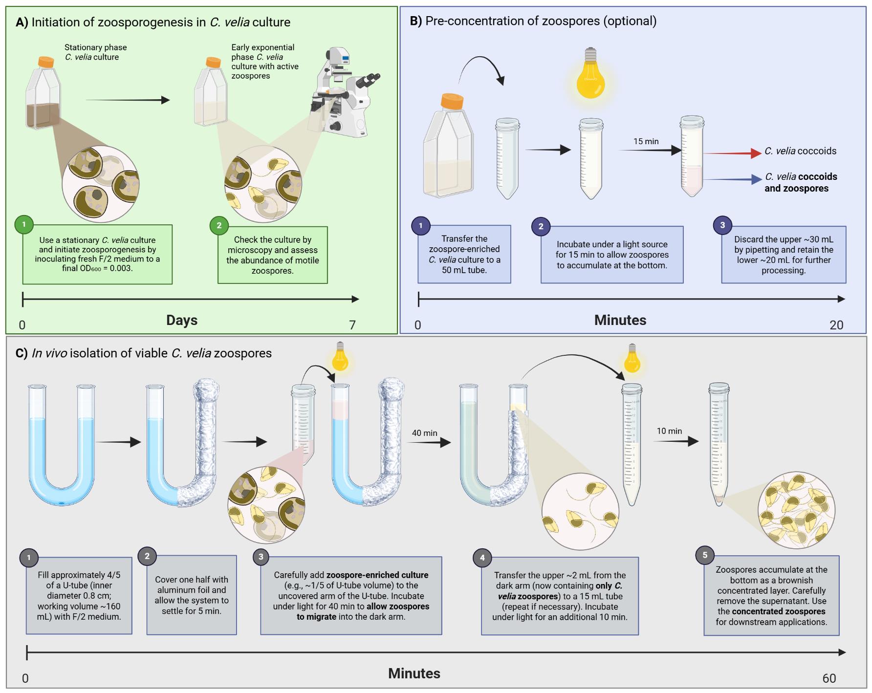
Example: For six U-tubes with an inner volume of 125 mL each, prepare 100 mL of F/2 medium per tube. Each U-tube will be topped with one-fifth of its volume (25 mL) of C. velia culture, with an additional 25 mL prepared as a backup reserve.
2. Maintain the culture under stationary conditions (no shaking) in a temperature-controlled incubator at 26 °C, under 100 μmol/m2/s illumination (verified with a lux meter) under a 12/12 h light/dark regime. (Optional) Check the culture daily by light microscopy for the presence of zoospores, 6 h after illumination.
3. On the seventh day (Figure 1A), 5 h after illumination, proceed with zoospore isolation according to the detailed steps in section A5.
Note: The zoospore-enriched fraction typically contains ≥400 zoospores per field of view (100× magnification) of motile zoospores, as estimated by light microscopy.
Critical: For efficient isolation, the zoospore-enriched culture should contain approximately ≥400 zoospores per field of view (100× magnification). Lower zoospore densities may significantly reduce isolation efficiency.
A5. Isolation of viable C. velia zoospores
0. (Optional) Pre-concentration:
a. Transfer the zoospore-enriched cultures into 50 mL centrifuge tubes.
b. Incubate the open 50 mL tubes under the light source for 15 min. Zoospores will begin accumulating at the conical tips of the tubes.
c. Discard, by pipetting, the upper 30 mL consisting of F/2 medium with floating C. velia coccoid cells.
d. Proceed to the next step with the remaining 20 mL containing C. velia zoospores and coccoids.
1. Prepare the U-tube system:
a. Light-proofing: Wrap one arm of the U-tube with aluminum foil to block light from entering that side. To maintain darkness, make a removable aluminum lid for the top of the wrapped arm. (See the graphical overview and Video 1 for details.)
b. Securing the U-tube: Place the U-tube in a stable transparent stand, ensuring both arms are vertical. A standard lab beaker is suitable.
c. Loading the medium: Fill a U-shaped glass tube with sterile F/2 medium, leaving the top 1/5 free to layer the C. velia culture.
Example: For a 125 mL total tube volume (50 cm long, inner diameter 0.8 cm), add 100 mL of F/2 medium, leaving 25 mL of free space.
d. Equilibration of the tube: Allow the sterile F/2 medium to settle undisturbed for 5 min.

2. Inoculation:
a. Thoroughly mix the C. velia culture containing active zoospores by gentle manual shaking of the cultivation flask.
b. Carefully pour the C. velia culture into the unwrapped arm of the U-tube alongside the sterile glass rod (see Video 1 for details). The added culture should occupy 1/5 of the U-tube volume.
Critical: Avoid moving or shaking the U-tube to prevent disturbing the medium. Do not pour the C. velia culture directly into the U-tube. The flow rate should allow the C. velia culture to proceed along the glass rod and down the inner wall of the U-tube, minimizing turbulence of the medium already present in the tube.
3. Illumination of the system:
a. Carefully place the U-tube under a white light source (380–720 nm) with an intensity of 100 μmol/m2/s (verified with a lux meter).
b. Ensure the light is directed only toward the unwrapped side of the U-tube.
c. Incubate the setup under light exposure for 40 min at room temperature (22–26 °C).
Critical: Maintain uniform illumination at 100 μmol/m2/s (verified with a lux meter), with a minimum distance of 15 cm between the light source and the upper part of the U-tube to ensure the system operates without disruption. A shield is recommended. Block the foil-covered side from ambient light to prevent directional bias; a consistent photophobic response is essential for efficient zoospore migration.
4. Collection of the isolated zoospores:
a. After 40 min, carefully remove the aluminum foil from the obscured arm of the U-tube.
b. Using a 5 mL pipette, gently move the pipette tip in a circular motion to withdraw 2 mL from the top layer of the previously obscured (foil-covered) arm.
c. Transfer this zoospore-enriched fraction into a transparent 15 mL centrifuge Falcon tube.
d. Repeat steps A5.4b–c if more U-tubes were used or if necessary.
Notes:
1. If you are using multiple U-tubes, combine the pooled 2 mL sample from step A5.4b into a single 15 mL centrifuge tube.
2. In cultures, where approximately 700–1,000 zoospores were counted per field of view (step A5.4c), a brownish cloud becomes visible within the upper 2 cm of the previously covered arm. The appearance of this cloud serves as a practical visual indicator of successful enrichment and corresponds to a zoospore-dominated fraction, as verified by light microscopy. If needed, you may check the zoospore-enriched fraction under an inverted microscope by transferring 100 μL into one well of a 96-well round-bottom plate (optional).
5. Concentration of the zoospores:
a. Place the 15 mL centrifuge tube containing the enriched fraction under a light source for 10 min to induce the accumulation of zoospores at the bottom of the tube (Figure 1B).
b. Carefully discard, by pipetting, the upper zoospore-free medium, retaining approximately 200 μL of medium.
Critical: A dark-brown layer may adhere to the wall of the glass U-tube or centrifuge tubes. Do not scrape it off, as it consists of freshly transformed C. velia coccoids. Zoospores accumulate at the bottom of the tube due to their photophobic behavior while remaining motile in the medium. In contrast, freshly formed coccoid cells adhere to the tube wall and are not resuspended under these conditions.
Note: The isolation procedure should be performed without interruption, as zoospores have a limited lifetime (up to ~8 h) [10]. Downstream handling depends on the intended application. Isolated zoospores can be used immediately as inoculum, fixed in 4% paraformaldehyde for microscopy-based analyses, or rapidly frozen in liquid nitrogen for downstream molecular analyses (e.g., -omics approaches).
B. Estimation of the average motility speed of C. velia zoospores
1. Prepare the zoospore culture as described in section A4.
2. On the day of the experiment, homogenize the culture (by gentle manual agitation) and quantify the zoospores by transferring 50 μL to well A1 of a 96-well round-bottom plate.
3. Fill the U-tube and track migration (detailed in section A5):
a. Load the zoospore-enriched culture into the unwrapped arm of a U-tube filled with F/2 medium.
b. Apply directional illumination to induce migration of the zoospores toward the dark arm as described in step A5.3a–c.
4. Time-course collection:
a. At 1-min intervals, transfer 50 μL of F/2 medium from the obscured U-tube arm into a well of a 96-well round-bottom plate. To do this, insert the pipette approximately 1 cm below the surface. Avoid pipetting from the same position each time.
b. Continue collecting one sample per minute for at least 40 min.
5. Analyze zoospore distribution:
a. Examine each well under 100× magnification (10× objective).
b. Check for the presence of zoospores in each well to determine migration over time.
c. Calculate the average swimming speed from the mean arrival time and the measured migration distance (length of the medium-filled U-tube).
Note: Perform this experiment in triplicate to ensure statistical robustness. This experiment can optionally be conducted in a dark room, with the unwrapped arm exposed to white light and the wrapped arm exposed to red light, to allow smooth operation without needing to uncover the open end of the U-tube every minute for time collection.
C. Estimation of the lifespan of isolated zoospores
1. Transfer freshly isolated zoospores:
a. Place 50 μL of zoospore-enriched medium into individual wells of a round-bottom 96-well plate.
Note: If necessary, dilute the zoospore suspension in F/2 medium to approximately 20 zoospores per well, or any other concentration that facilitates accurate counting.
b. Prepare at least three biological replicates.
2. Temporal observation:
a. At 30-min intervals, observe each well under an inverted microscope (100×).
b. Count motile (actively swimming) and non-motile (coccoid) cells.
Critical: Maintain the temperature at 26 °C and avoid direct light exposure between observations to mimic culture conditions.
3. Record endpoint:
a. Continue observations until 100% transformation to the coccoid stage is achieved (approximately 8 h) [10].
b. Estimate the average lifespan as the midpoint between observation of motile zoospores and the point at which all cells have completed transformation into non-motile forms.
Note: Waste disposal: At the end of the protocol, and before disposal, decontaminate the residual medium in the U-shaped tubes containing the coccoid cells of C. velia, along with any remaining C. velia culture, with 1% (v/v) bleach solution [prepared by diluting commercial bleach stock, ~3%–5% (w/v) sodium hypochlorite, 1:5 in distilled water].
Validation of protocol
The directional light–based isolation of C. velia zoospores has been validated by repeated independent experiments (n ≥ 3). After 40 min of directional illumination, zoospores reproducibly migrate into the dark arm of the U-tube, resulting in a zoospore-enriched fraction with no coccoid stages present, as confirmed by microscopic examination.
This protocol, or parts of it, has been used and validated in the following research article: Richtová et al. [10]. Circadian rhythms and circadian clock gene homologs of complex alga Chromera velia. Frontiers in Plant Science 14:1226027 (see Section 2.3; Figure 2; Supplementary Figure 3).
General notes and troubleshooting
General notes
1. Zoosporogenesis in C. velia is highly sensitive to light quality. A blue-enriched spectrum (420–460 nm) is critical for reliable induction and isolation efficiency. Variations in light source or intensity may significantly affect zoospore production.
2. The physiological state of the culture strongly influences isolation success. Stable cultivation under consistent light/dark conditions improves reproducibility. Cultures recently stressed, overgrown, or contaminated may exhibit reduced or delayed zoosporogenesis.
3. The initial inoculum density (OD600 = 0.003–0.004, measured using a spectrometer) is critical. Higher densities lead to increased culture turbidity and a higher proportion of coccoid stages, thereby reducing migration efficiency (see Section A).
4. The isolation method relies on active negative phototaxis. Proceeding with isolation outside the peak of zoosporogenesis may result in significantly lower yields of isolated zoospores.
5. This protocol may be adapted for other flagellated protists exhibiting light-responsive motility; however, light intensity, incubation time, and tube dimensions must be empirically optimized.
6. The use of a U-shaped glass tube is essential for establishing a stable light gradient and enabling directional migration of zoospores. The protocol can also be performed using smaller tubes with similar geometry; however, reduced tube dimensions (e.g., lower volume or smaller diameter) result in proportionally lower yields of isolated zoospores.
Troubleshooting
Problem 1: No zoospores in the culture after 7 days (related to Recipes section and steps A1.1–4)
Possible cause: Composition of the F/2 medium. C. velia zoosporogenesis is highly sensitive to the presence of key elements in the F/2 medium. The complete absence of a nitrate source can block zoosporogenesis. The complete absence of phosphate results in delayed and weaker zoosporogenesis.
Solution: Prepare new F/2 medium.
Problem 2: No zoospores in the culture after 7 days (related to section A, culture maintenance).
Possible cause: Fungal infection in the culture.
Solutions: Examine the culture under a microscope for fungal spores or infected cells. Treat the culture by adding fresh Azoxystrobin into the F/2 medium. Maintain the culture in F/2 Azo for one week, then use it to inoculate a new culture in F/2 Azo.
Problem 3: No zoospores after isolation (related to section A5, loading the U-tube).
Possible causes: The chosen culture was too dense. No zoospore-enriched culture of C. velia is free of coccoid cells. Coccoid cells present in the culture will settle and accumulate at the bottom of the U-tube, forming a natural shade where zoospores will hide from the light source.
Solution: Dilute the original culture.
Problem 4: No zoospores after isolation (related to sections A3–A4, verification of zoospore abundance before isolation).
Possible cause: Not enough zoospores in the culture. We recommend not proceeding with isolation if the number of zoospores per field of view is lower than 200; the optimal number for isolation is ≥400 zoospores per field of view (100× magnification).
Solution: Inoculate a new culture. Make sure you are also not experiencing the abovementioned problems 1 or 2.
Problem 5: Zoospores are present but fail to migrate (related to section A5, directional light exposure and migration step).
Possible causes: Inadequate light conditions in terms of intensity, spectrum, or direction reduce motility of zoospores due to suboptimal physiological state, or excessive culture density leading to self-shading effects within the U-tube (see Problem 3). The isolation relies on active negative phototaxis, which may be impaired under these conditions.
Solutions: Ensure the use of a white or blue-enriched light source (~100 μmol/m2/s) and correct positioning relative to the U-tube (see section A5). Verify that the culture contains actively motile zoospores and is used at the appropriate density. If the culture is too dense, dilute prior to loading (see Problem 3).
Problem 6: Mixed populations after isolation (related to section A5, sampling from the U-tube).
Possible cause: The sample was collected too close to the middle/lower part of the wrapped arm, where coccoid cells may settle or remain in suspension. This can result in the co-collection of non-motile stages together with zoospores.
Solution: To define a safe zoospore collecting zone, do not insert the pipette deeper than the volume corresponding to the upper 1/5 of the total U-tube volume from the foil-covered side. For example, in a U-tube with a total volume of 100 mL, collect only from the upper 20 mL region of the foil-covered arm. Avoid sampling near the middle of the U-tube.
Problem 7: Contamination introduced during U-tube handling (related to section A5, handling and transfer steps).
Possible cause: Exposure of the open U-tube system to environmental contaminants (e.g., airborne particles, microorganisms) during handling.
Solution: Although the protocol is not designed to be fully sterile, it is recommended to perform the procedure in a clean working environment and to minimize unnecessary exposure of the U-tube openings. Use clean pipette tips and freshly prepared medium to limit the introduction of contaminants that may interfere with microscopic observation or downstream applications. If sterile conditions are required, the entire setup can be handled in a laminar flow hood.
Supplementary information
The following supporting information can be downloaded here:
1. Figure S1. Technical drawing of the U-shaped glass tube used for zoospore isolation.
Acknowledgments
Author’s contribution: Conceptualization, J.R.; Investigation, J.R., D.E.; Writing—Original Draft, J.R., D.E.; Visualization (Graphical overview, figures, video), J.R.; Writing—Review & Editing, J.R., D.E., M.O.; Funding acquisition, M.O.; Supervision, M.O.
This work was supported by the Czech Science Foundation grant no 25-15611S (M.O.).
This protocol is adapted from the original paper [10].
We thank Matěj Pecha for filming and video processing.
The following figures were created using BioRender: Graphical overview: Richtová, J. (2026) https://BioRender.com/141rgbx.
Competing interests
The authors declare no conflicts of interest.
References
- Cavalier-Smith, T. (2018). Kingdom Chromista and its eight phyla: a new synthesis emphasising periplastid protein targeting, cytoskeletal and periplastid evolution, and ancient divergences. Protoplasma. 255(1): 297–357. https://doi.org/10.1007/s00709-017-1147-3
- Moore, R. B., Oborník, M., Janouškovec, J., Chrudimský, T., Vancová, M., Green, D. H., Wright, S. W., Davies, N. W., Bolch, C. J. S., Heimann, K., et al. (2008). A photosynthetic alveolate closely related to apicomplexan parasites. Nature. 451(7181): 959–963. https://doi.org/10.1038/nature06635
- Cumbo, V. R., Baird, A. H., Moore, R. B., Negri, A. P., Neilan, B. A., Salih, A., van Oppen, M. J. H., Wang, Y. and Marquis, C. P. (2013). Chromera velia is endosymbiotic in larvae of the reef corals Acropora digitifera and A. tenuis. Protist. 164(2): 237–244. https://doi.org/10.1016/j.protis.2012.08.003
- Mohamed, A. R., Cumbo, V. R., Harii, S., Shinzato, C., Chan, C. X., Ragan, M. A., Satoh, N., Ball, E. E. and Miller, D. J. (2018). Deciphering the nature of the coral-Chromera association. ISME J. 12(3): 776–790. https://doi.org/10.1038/s41396-017-0005-9
- Oborník, M., Kručinská, J. and Esson, H. (2016). Life cycles of chromerids resemble those of colpodellids and apicomplexan parasites. Perspect Phycol. 3(1): 21–27. https://doi.org/10.1127/pip/2016/0038
- Oborník, M., Vancová, M., Lai, D. H., Janouškovec, J., Keeling, P. J. and Lukeš, J. (2011). Morphology and ultrastructure of multiple life cycle stages of the photosynthetic relative of Apicomplexa, Chromera velia. Protist. 162(1): 115–130. https://doi.org/10.1016/j.protis.2010.02.004
- Portman, N., Foster, C., Walker, G. and Šlapetaa, J. (2014). Evidence of intraflagellar transport and apical complex formation in a free-living relative of the apicomplexa. Eukaryot Cell. 13(1): 10–20. https://doi.org/10.1128/EC.00155-13
- Oborník, M. (2020). Photoparasitism as an Intermediate State in the Evolution of Apicomplexan Parasites. In Trends Parasitol. (Vol. 36, Issue 9, pp. 727–734). Elsevier Ltd. https://doi.org/10.1016/j.pt.2020.06.002
- Agrawal, S. C. (2012). Factors controlling induction of reproduction in algae-review: The text. Folia Microbiol. (Praha). 57(5): 387–407. https://doi.org/10.1007/s12223-012-0147-0
- Richtová, J., Bazalová, O., Horák, A., Tomčala, A., Gonepogu, V. G., Oborník, M. and Doležel, D. (2023). Circadian rhythms and circadian clock gene homologs of complex alga Chromera velia. Front Plant Sci. 14. https://doi.org/10.3389/fpls.2023.1226027
- Amon, J. P. and French, K. H. (2004). Photoresponses of the marine protist Ulkenia sp. zoospores to ambient, artificial and bioluminescent light. Mycologia. 96(3): 463–469. https://doi.org/10.1080/15572536.2005.11832945
- Swafford, A. J. M. and Oakley, T. H. (2018). Multimodal sensorimotor system in unicellular zoospores of a fungus. J Exp Biol. 221(2). https://doi.org/10.1242/jeb.163196
- Callow, M. E. and Callow, J. A. (2000). Substratum location and zoospore behaviour in the fouling alga Enteromorpha. Biofouling. 15(1–3): 49–56. https://doi.org/10.1080/08927010009386297
- Maier, M. A., Uchii, K., Peterson, T. D. and Kagami, M. (2016). Evaluation of daphnid grazing on microscopic zoosporic fungi by using comparative threshold cycle quantitative PCR. Appl Environ Microbiol. 82(13): 3868–3874. https://doi.org/10.1128/AEM.00087-16
- Penaud, V., Alahmad, A., De Vrieze, M., Bouteiller, M., Eude, M., Bernardon-Mery, A., Trinsoutrot-Gattin, I., Laval, K. and Gauthier, A. (2025). In vitro biocontrol potential of plant extract-based formulation against infection structures of Phytophthora infestans along with lower non-target effects. Front Microbiol. 16. https://doi.org/10.3389/fmicb.2025.1569281
- Hiraizumi, M. and Tagawa, Y. (2014). Isolation and characterization of flagellar filament from zoospores of Dermatophilus congolensis. Vet Microbiol. 173(1–2): 141–146. https://doi.org/10.1016/j.vetmic.2014.07.017
- Tomley, F. (1997). Techniques for Isolation and Characterization of Apical Organelles from Eimeria tenella Sporozoites. In METHODS: A Companion to Methods in Enzymology (Vol. 13).
Article Information
Publication history
Received: Mar 6, 2026
Accepted: Jun 4, 2026
Available online: Jun 24, 2026
Published: Jul 20, 2026
Copyright
© 2026 The Author(s); This is an open access article under the CC BY-NC license (https://creativecommons.org/licenses/by-nc/4.0/).
How to cite
Richtová, J., Ennaceur, D. and Oborník, M. (2026). Separating Chromera velia Zoospores From Culture and Estimating Their Average Motility Speed and Lifespan. Bio-protocol 16(14): e5756. DOI: 10.21769/BioProtoc.5756.

 Download Citation in RIS Format
Category
Microbiology > Microbial cell biology > Cell isolation and culture
Microbiology > Microbial physiology > Phototaxis
Plant Science > Phycology > Cell isolation and culture
Do you have any questions about this protocol?
Post your question to gather feedback from the community. We will also invite the authors of this article to respond.
 Post a Question


