


- Protocols

- Articles and Issues
- For Authors
- About
- Become a Reviewer


Extraction and Isolation of Extracellular Vesicles From Piper betle Leaves Using the Apoplastic Fluid Washing and Size Exclusion Chromatography Method


Published: Vol 16, Iss 9, May 5, 2026 DOI: 10.21769/BioProtoc.5685 Views: 619
Reviewed by: Pawan KumarMarisa ConteAnonymous reviewer(s)
 PDF
PDF  Ask a question
Original research article
The authors used this protocol in:

Nov 2024
Advertisement



Protocol Collections
Comprehensive collections of detailed, peer-reviewed protocols focusing on specific topics




Related protocols
Abstract
Plant-derived extracellular vesicles (PDEVs) have emerged as important mediators of intercellular communication and hold growing potential in therapeutic applications. However, standardized methods for their isolation, particularly from Piper betle leaves (PBL), remain unexplored. Existing apoplastic fluid washing (AFW) extraction techniques typically rely on manual syringe infiltration, which often leads to inconsistent pressure control, variable yields, and increased risk of tissue damage. This protocol describes a vacuum-assisted AFW extraction method optimized for the recovery of intact extracellular vesicles (EVs) from PBL. The workflow features controlled negative pressure using a vacuum pump and chamber to achieve more efficient leaf infiltration compared to infiltration using the syringe method and reproducible apoplastic fluid (AF) collection with subsequent low-speed centrifugation steps, to ensure minimal contamination and preservation of vesicle integrity. Piper betle–derived extracellular vesicle (PBdEV) isolation and purification steps are performed using size exclusion chromatography (SEC). The size and concentration of PBdEVs were confirmed using nanoparticle tracking analysis (NTA), whereas the cup-shaped and lipid bilayer morphology of the EVs were confirmed using transmission electron microscopy (TEM). The method is scalable and adaptable to various leaf morphologies and physiological states, making it suitable for both exploratory and high-throughput studies. Overall, this protocol provides a more consistent, efficient, and tissue-preserving alternative to traditional syringe-based AF extraction methods, offering higher-quality EV preparations for plant EV research.
Key features
• This protocol focuses on the extraction of Piper betle–derived extracellular vesicles (PBdEVs) using a gentle approach to maintain the vesicles’ morphology.
• This protocol is suitable for large-scale experiments with multiple biological replicates or different leaf samples of similar morphology.
Keywords: Apoplastic fluid washing Piper betle leaves
Extracellular vesicles
Piper betle–derived extracellular vesicles
Piper betle leaves
Extracellular vesicles
Piper betle–derived extracellular vesicles
Graphical overview
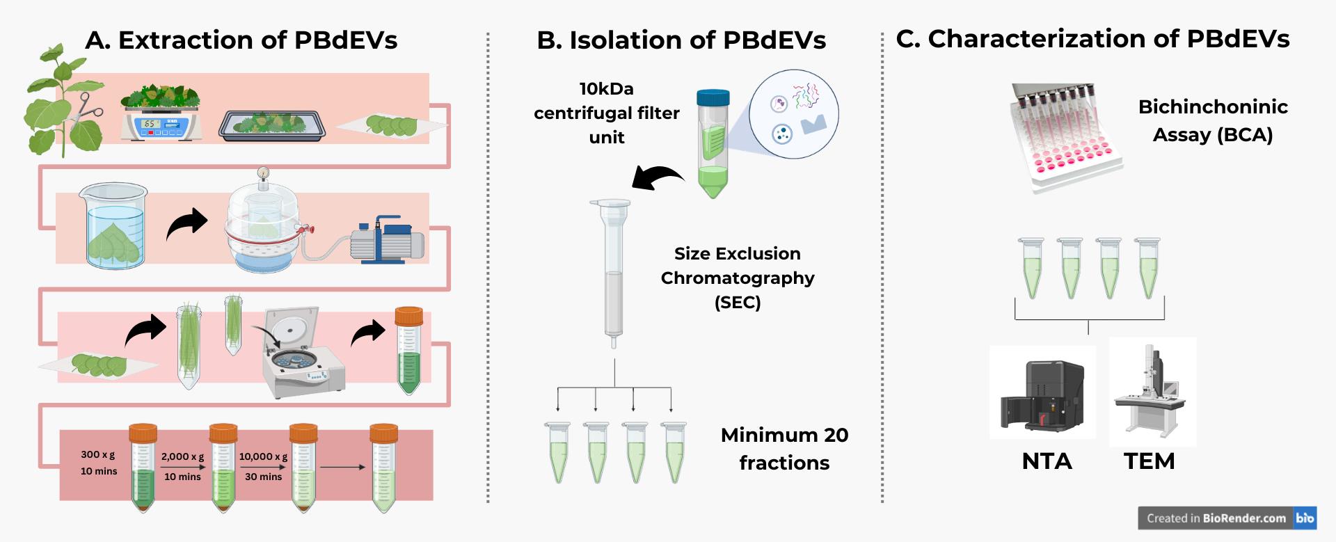
Overview of the workflow to extract and isolate Piper betle–derived extracellular vesicles (PBdEVs). The complete workflow of extracting, isolating, and characterizing PBdEVs involves (A) extracting the PBdEVs using the apoplastic fluid washing (AFW) method and (B) applying size exclusion chromatography (SEC) to gently isolate PBdEVs. After that, (C) bicinchoninic assay (BCA) is performed to determine the protein-to-particle ratio, before identification of the isolated particles using nanoparticle tracking analysis (NTA) and transmission electron microscopy (TEM). The identification methods are not included in this protocol.
Background
Extracellular vesicles (EVs) are nanoscale, membrane-bound particles released by both animal and plant cells [1]. In plants, EVs mediate intercellular communication, participate in defense responses, and transport bioactive cargoes such as proteins, lipids, nucleic acids, and small metabolites [2] across cell walls into the apoplast [3]. Plant-derived EVs are gaining popularity due to their roles in interspecies communication [4], potential application in nanoscale drug delivery, and disrupting oncogenic communication [5–8]. Several methods have been developed to isolate plant EVs, including differential ultracentrifugation, density gradient separation, and precipitation-based approaches [9–11]. While these methods have enabled biochemical characterization and functional studies, they often face challenges such as low yield, co-isolation of contaminants, the requirement of specialized equipment, or limited applicability to different plant tissues [12]. Apoplastic fluid washing (AFW) is a proposed method for harvesting plant extracellular contents with minimal intracellular contamination [13–15], making it well-suited for the isolation of EVs for biochemical characterization and functional testing. Due to their richness in secondary metabolites exhibiting antibacterial and anti-inflammatory activities [16,17] and their abundance in Malaysia [18], Piper betle leaves represent a promising source of plant-derived EVs for further exploration of their therapeutic potential [19–21].
Materials and reagents
Biological materials
1. Piper betle leaves from the family Piperaceae; young to mature leaves, aged 20-30 days old
Note: Young to mature Piper betle leaves have light to deep green–colored leaves (refer to Figure 1A, B), besides being soft to the touch instead of feathery, and can be bent without breaking. The reason why age matters in choosing the leaves is because of the integrity level of the cell wall. In young to mature leaves, cell walls are less developed, thus better in permitting the infiltration of buffer with gentle pressure.
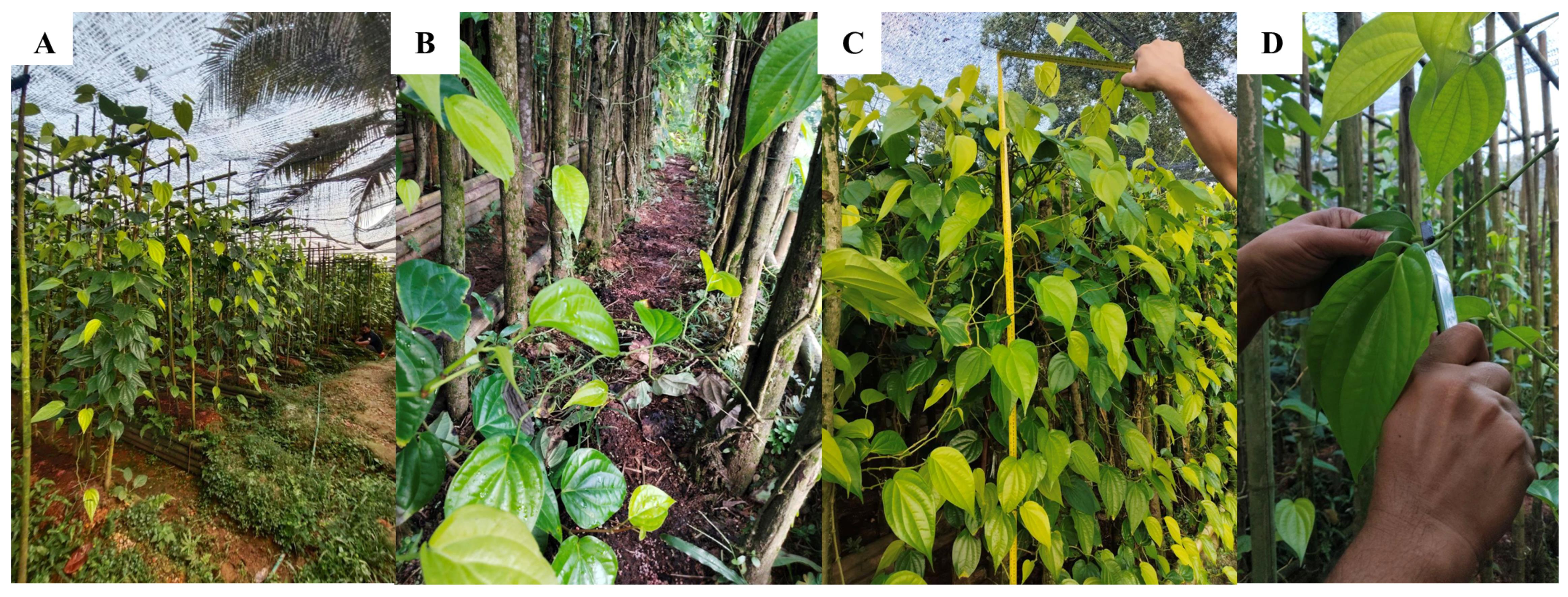
Figure 1. Harvesting Piper betle leaves. (A) Propagation and cultivation conditions of Piper betle. The plant is propagated vegetatively using stem cuttings containing 2–3 nodes, planted on a raised bed at 60–90 cm spacing with 2 m of supporting pole for climbing, and grown under partial shade of the black net. (B) The Piper betle is supplied with NPK fertilizer to support healthy leaf growth. (C) The plant can reach up to 2 m high after 6 months, when it is ready to be harvested. (D) The farmer is instructed to cut the desired leaves at the petiole with a clean knife.
Reagents
1. Phosphate-buffered saline (PBS), pH 7.4 (Biomedia, catalog number: 820301-1); store at 4 °C
2. 70% ethanol (EtOH) (Chemiz, CAS number: 64-17-5); store at room temperature (RT) in a flammable liquid storage cupboard
Caution: This chemical is flammable.
3. SepharoseTM CL-2B (Cytiva Sweden AB, catalog number: 10346215); store at RT away from direct sunlight and heat
Caution: This chemical is flammable.
4. Milli-Q (Merck Millipore); keep at 4 °C for the purpose of this protocol; store at RT if not used
Laboratory supplies
1. Glass chamber A (height: 220 mm; diameter: 190 mm; thickness: 5 mm) (PLT Scientific Sdn Bhd)
Note: This glass chamber was ordered and modified based on our needs. You may need a bigger or smaller chamber depending on the size of your leaves, vacuum chamber, vacuum pump, and experiment scale.
2. Glass chamber B (height: 200 mm; diameter: 150 mm; thickness: 5 mm) (PLT Scientific Sdn Bhd)
Note: This glass chamber was ordered and modified based on our needs. You may need a bigger or smaller chamber depending on the size of your leaves, vacuum chamber, vacuum pump, and experiment scale.
3. Parafilm M All-Purpose Laboratory Film (American Can, catalog number: LMA-PM996)
4. 50 mL conical centrifuge tubes (Biomedia, catalog number: 940204)
5. 20 mL needleless syringe (Terumo, catalog number: 250203B)
6. Pipette tips, 200 and 1,000 μL
7. Econo-Pac® Chromatography Columns kit (Bio-Rad, catalog number: 7321010)
8. Serological pipette, 10 and 25 mL (Bio-Rad, catalog numbers: 031024CM09, 051922CM08)
9. Blunt forceps (LLG Labware, catalog number: 4008476)
10. 500 mL glass beaker (waste container)
11. Cling wrap (Analisa Resources, catalog number: 39201025)
12. Amicon® Ultra Centrifugal Filter, 10 kDa MWCO (Millipore, catalog number: UFC9010)
13. Eppendorf Safe-Lock® tubes biobased 1.5 mL (Eppendorf, catalog number: 0030123093)
Equipment
1. Vacuum chamber and pump (HEXO, model: RS-1.5)
Note: The pump oil may not be provided and needs to be bought separately, if not provided by the manufacturer.
2. Refrigerated centrifuge (Centrifuge 5804 R) (Eppendorf, catalog number: 5805FM568504) (fixed-angle centrifuge)
3. Tabletop centrifuge (Trabota, model: J74276-M000) (fixed-angle centrifuge)
4. Micropipette 20–200 μL (Joanlab, model: 01235556)
5. Micropipette 100–1,000 μL (Joanlab, model: 01242536)
6. Analytical laboratory scale (OHAUS, model: PA214)
7. Freezer (-80 °C)
8. Retort stand or box with holes
9. Electronic pipette with stand (Joanlab, model: EP100Pro)
10. Vegetable washing basket (KECH, catalog number: 1079134)
Procedure
A. Collection of Piper betle leaves
1. Cut healthy and mature betel leaves at the petiole using a clean, sharp knife. Mature betel leaves are light to deep green in color and have a wide diameter (approximately 7–9 cm) and length (approximately 13–15 cm).
Note: Betel plants were not cultivated by the researchers. Instead, leaves were purchased from a betel farm in Bukit Beruntung, Rawang, Selangor, Malaysia (coordinates: N 03°26′2.67″, E 101°33′4.543″). However, the collection process was observed by the researchers, and specific instructions regarding the collection protocol were provided to the farmer.
2. Wash the leaves under clean running water to remove surface dirt. Subsequently, gently arrange the leaves in a box or container layered with moistened tissues or cloth over and between the leaves to maintain freshness and reduce water stress during transportation.
Critical: Transportation time should be kept to a minimum (<30–45 min), and leaves should be processed as soon as possible to ensure maximal freshness during the PBdEV isolation process.
B. Extraction of apoplast washing fluid
1. Wash the leaves gently again under running water. Disinfect the leaves by briefly soaking them in 70% ethanol for 30 s to 1 min to remove surface contaminants. After washing and disinfection, allow the leaves to air-dry on clean tissues. Weigh the leaves to obtain the dry weight before infiltration and record the values in the data table (Table 1).
Critical: Do not prolong the 70% ethanol disinfection step, as excessive exposure may disrupt cellular integrity and lead to organelle contamination.
Table 1. Recording the dry weight and wet weight. The batch number is associated with its dry weight (before infiltration), wet weight (after infiltration), the estimated infiltrated buffer, and the apoplastic fluid, obtained by subtracting the dry weight from the wet weight (1 g equals 1 mL of buffer).
| Batch | Dry weight (g) | Wet weight (g) | Est. infiltrated buffer (mL) | Obtained apoplastic fluid (mL) |
|---|---|---|---|---|
| 1 | 501.93 | 536.50 | 34.57 | 33.50 |
| 2 | 511.08 | 562.58 | 51.50 | 51.00 |
| 3 | 509.61 | 555.81 | 46.20 | 44.00 |
| 4 | 527.52 | 591.69 | 64.17 | 63.66 |
2. Arrange the leaves in a larger-diameter glass beaker (glass beaker A) with the abaxial (lower) surface facing upward (Figure 2A). Pour cold PBS into glass beaker A until the leaves are fully submerged. Place a smaller-diameter glass beaker (glass beaker B) inside glass beaker A to apply gentle pressure to the leaves (Figure 2C).
Note: This setup prevents the leaves from floating during the vacuuming process and avoids infiltration failure.
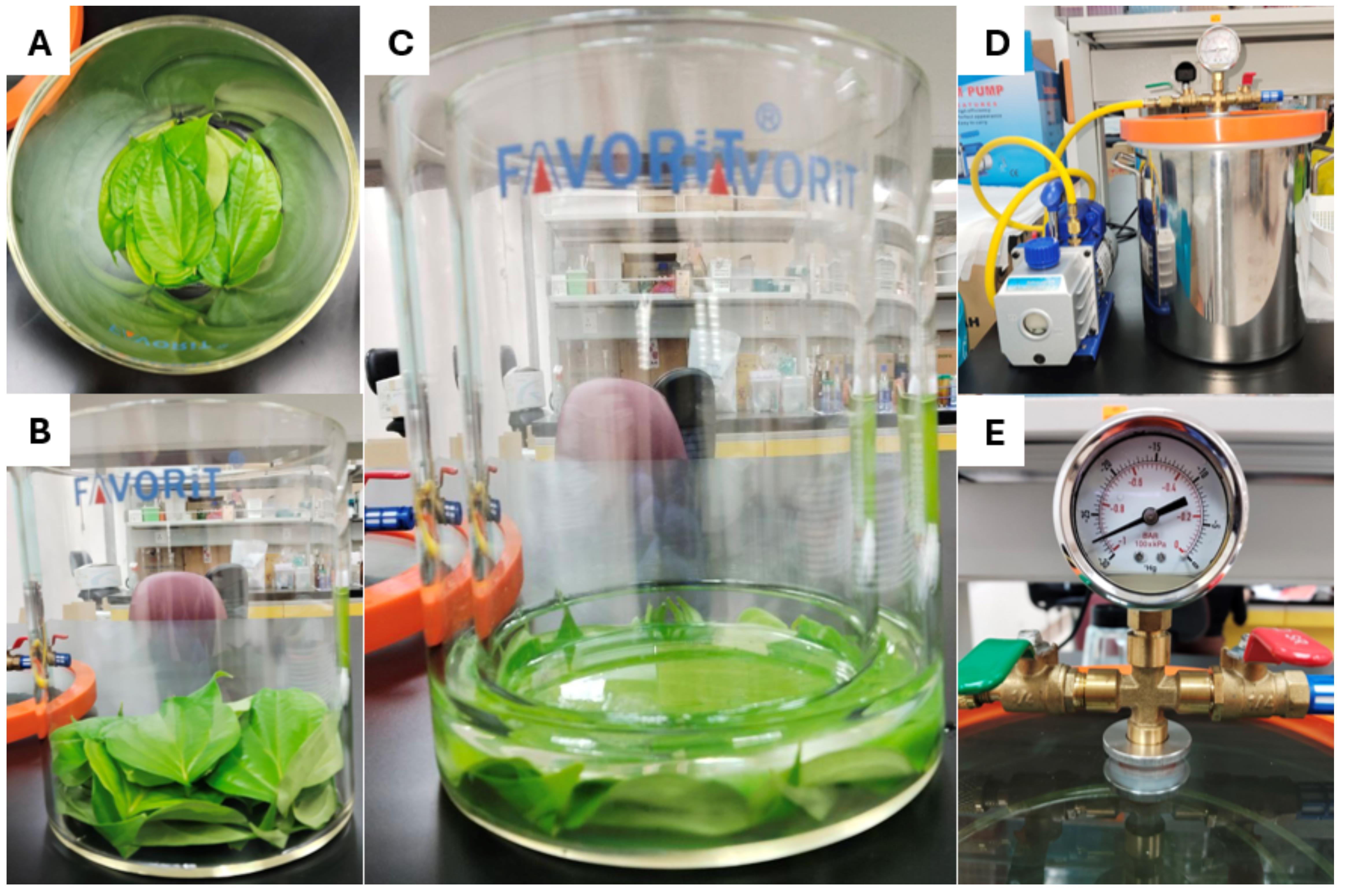
Figure 2. Infiltration protocol equipment and setup. (A) Top view of the leaf’s orientation in glass chamber A, where the abaxial side is facing upward. (B) Side view of the leaf orientation in glass beaker A. (C) Side view of glass beaker B pressing on the leaves in glass beaker A. (D) Vacuum pump and vacuum chamber setup (see Equipment). (E) Gauge showing the lowest pressure that the vacuum pump can achieve.
3. Carefully place the assembled setup inside the vacuum chamber. Connect the hoses to the vacuum pump and pressure gauge and close the outlet valve (Figure 2D).
4. Turn on the vacuum pump and monitor the reading on the gauge. Once the pressure reaches between -20 and -30 kPa (Figure 2E), close the inlet valve and gradually open the outlet valve by approximately 3 cm to slowly introduce air into the chamber. Observe changes in leaf appearance through the glass lid. The appearance of dark spots on the leaves indicates buffer infiltration (Figure 3C).
Caution: Repeat the infiltration step up to four times if the initial infiltration efficiency is unsatisfactory (<80%). Limit the number of repetitions to minimize the risk of cell rupture and the release of intracellular contaminants into the apoplastic fluid (AF).
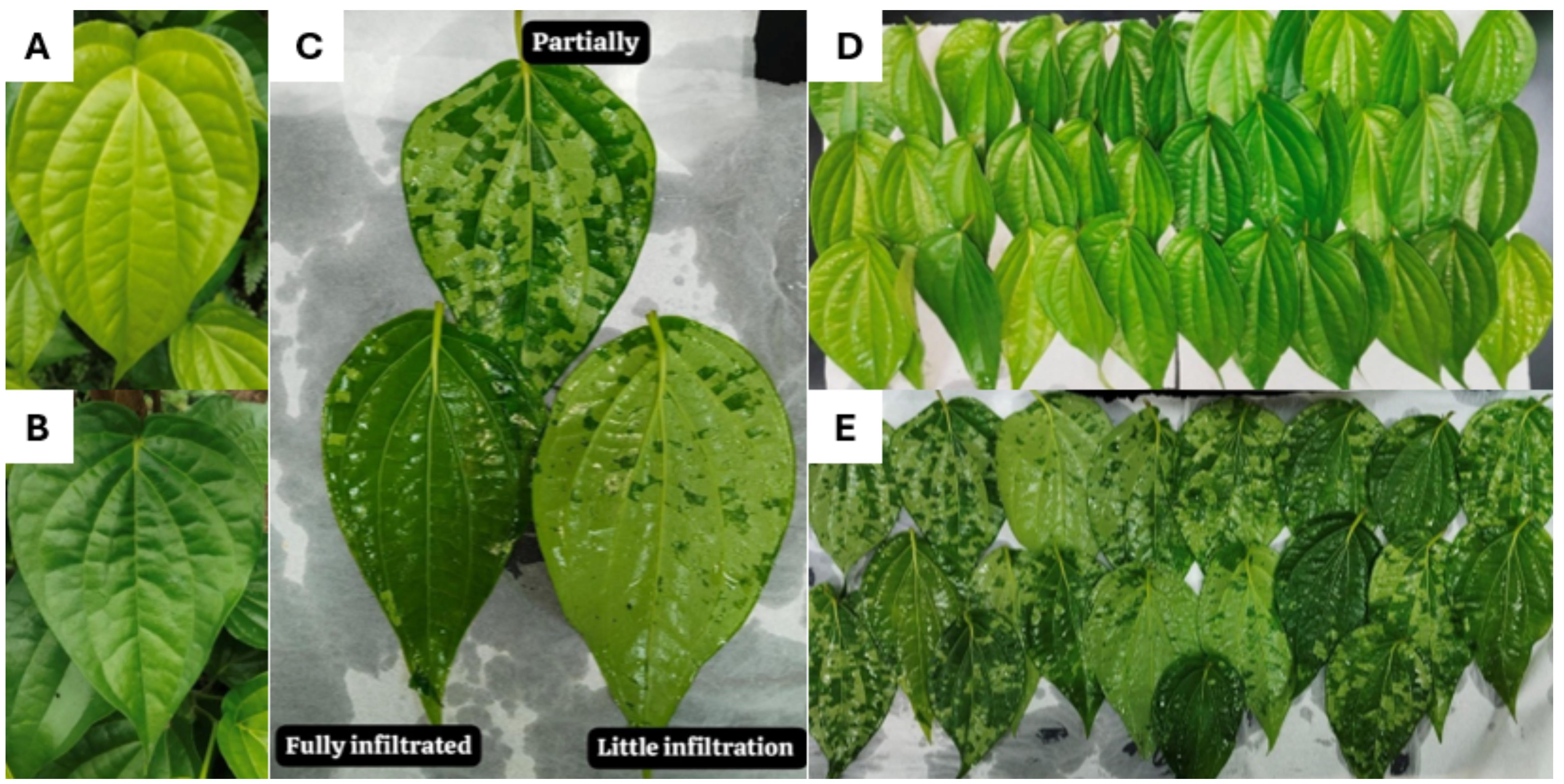
Figure 3. Conditions of the leaves. (A) Young betel leaves, with light green color appearance. (B) Older betel leaves, with deep green color and a more leathery appearance. (C) Rate of infiltration assessment and observation. Satisfactory infiltration is shown as the leaves with the fully infiltrated label (>80% infiltration rate). (D) Betel leaves after washing and before infiltration. (E) Betel leaves after infiltration, air-drying on top of clean tissues.
5. After infiltration, carefully take out the leaves from the buffer and lay them out on a clean tissue to dry. Gently pat the leaves to remove excess buffers from the surface. Weigh the leaves to get their wet weight and record the values in the data table (Table 1).
6. Once the leaves are dried, divide them into batches, with each batch containing five leaves of similar sizes and comparable infiltration efficiency. Roll the leaves together using cling wrap or parafilm. Cut a piece of cling wrap or parafilm large enough to accommodate all five leaves. Slightly overlap the leaves on the wrap and roll them tightly to form a compact bundle that fits into the barrel of a 20 mL needleless syringe (Figure 4A).
7. Remove the plunger from the 20 mL needless syringe. Insert the roll into the barrel and then insert the barrel into a 50 mL conical centrifuge tube and secure it with parafilm (Figure 4B). Prepare each batch promptly to avoid inducing plant cell stress. Place completed setups on ice while preparing subsequent batches to maintain a low internal buffer temperature.
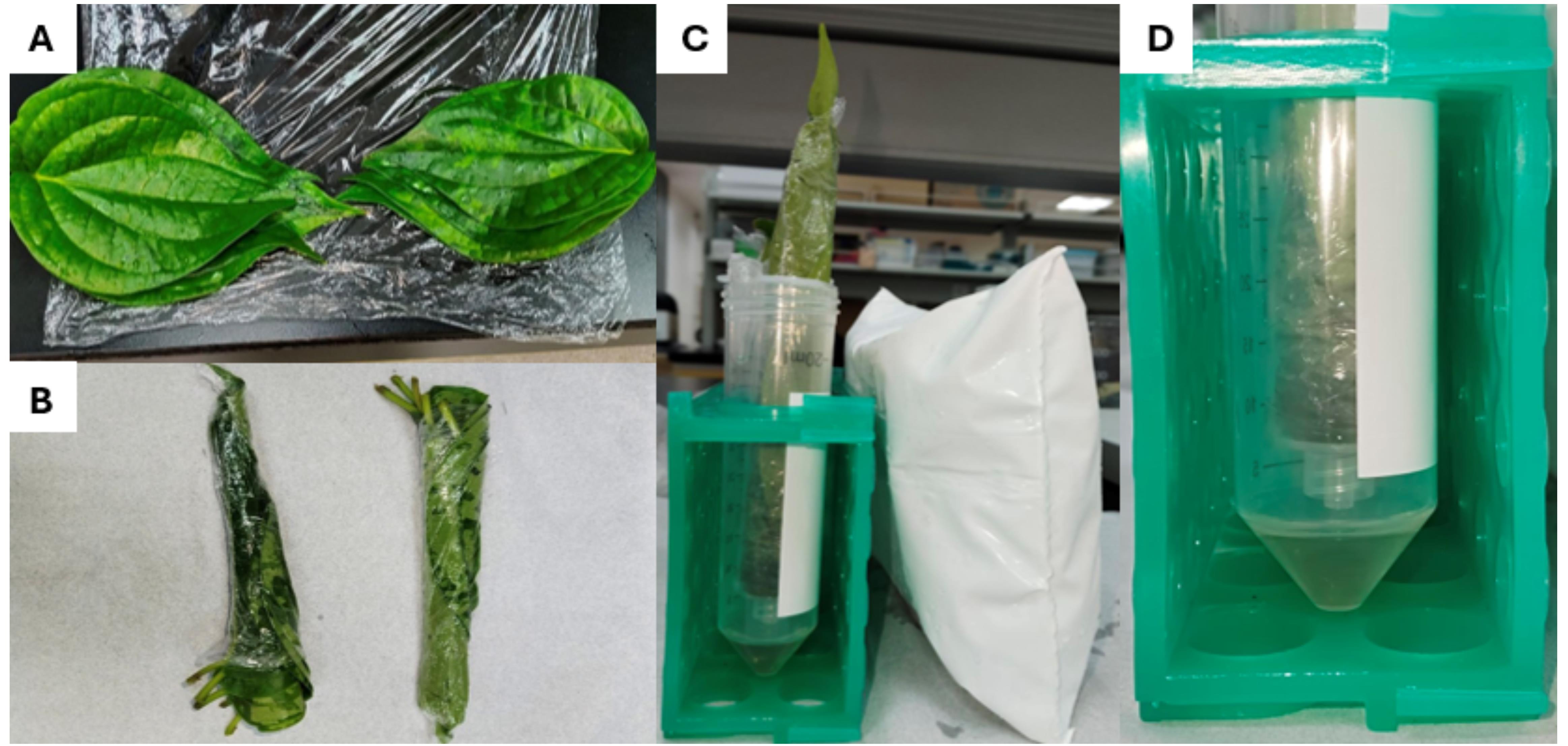
Figure 4. Apoplastic fluid collection protocol. (A) Infiltrated leaves arranged on top of each other on a piece of cling wrap, ready to be rolled. (B) Rolled up betel leaves, where one end is left open for the apoplastic fluid (AF) to flow out. (C) Rolled up betel leaves placed in the barrel of a 20 mL needleless syringe. (D) AF collected in the bottom of the centrifuge tube after centrifugation at 1,000× g for 20 min.
8. Once ready, centrifuge the conical centrifuge tubes at 1,000× g for 20 min to collect the AF. The AF collects at the bottom of the conical centrifuge tube. Transfer the AF into a clean conical centrifuge tube and keep it on ice.
9. After collecting AF from all batches, perform differential centrifugation as follows: a) 300× g for 10 min, b) 2,000× g for 10 min, and c) 10,000× g for 30 min. After each centrifugation step, transfer the supernatant into a new clean conical centrifuge tube and discard the pellet.
10. Store the clean AF in -80 °C until further use. Minimize freeze-thaw cycles to reduce the risk of rupturing the vesicle membrane or aggregation of the EVs.
Pause point: This step marks the end of day 1. Ensure that the AF is cleared of large cellular debris and contaminants before long-term storage at -80 °C.
C. Concentrating the AF for SEC
1. Load the clarified apoplastic fluid batch by batch into the 10 kDa centrifugal filter unit (CFU) and centrifuge at 3,500× g for 20 min until all sample is concentrated to an end volume of 500 μL.
Note: Since the CFU can only fit 15 mL of sample at once, four CFUs may be used to expedite the concentrating process.
Critical: When there is less than 15 mL of sample left, the centrifuge speed and time should be reduced suitably to the amount of sample left. The end volume of the sample should be approximately 500 μL.
2. In between centrifugation, gently agitate the sample by pipetting the sample up and down between the filters. This is done to ensure that minimal loose particles are stuck to the filters, which would prevent effective filtration.
3. Transfer the 500 μL of concentrated sample into an Eppendorf tube and keep at -80 °C until further use.
D. Isolation of PBdEVs using the SEC method
1. Prepare an empty chromatography column (Econo-Pac® Chromatography Columns) by securing it in an upright position using a retort stand or a custom stand (e.g., a box with holes) (Figure 5A).
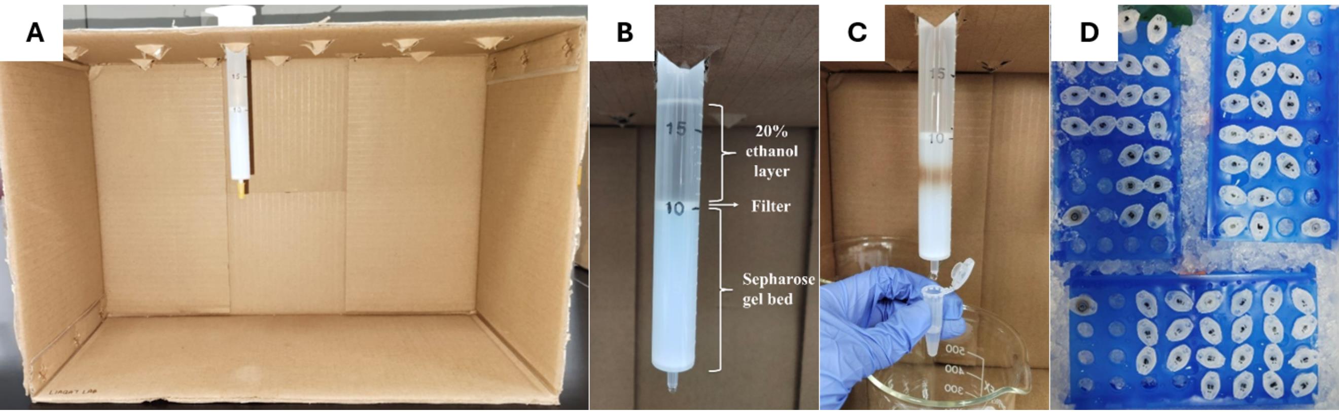
Figure 5. Size exclusion chromatography (SEC) isolation method to isolate Piper betle–derived extracellular vesicles (PBdEVs). (A) An innovation of a box stand to hold many chromatography columns at once to facilitate observation and the collection process. (B) Layers of 20% ethanol and Sepharose gel bed, separated by a filter that is inserted at a straight orientation to compact the gel bed to the 10 mL meniscus. (C) Fraction collection process, as the sample (brown layer) is moving through the Sepharose gel bed. (D) Collected fractions on ice in labeled 1.5 mL Eppendorf tubes.
2. Vigorously shake or mix the gel (Sepharose CL-2B) until the gel beads and ethanol are fully homogenized, and no distinct layers are observed.
Note: The 20% ethanol in the solution preserves the integrity of the gel beads. When left undisturbed, the ethanol separates from the beads due to differences in density.
3. Load 15 mL of the Sepharose CL-2B gel into the column using a serological pipette. Allow the gel to settle at RT with the cap loosened for at least 2 h, during which the gel beads and 20% ethanol separate into distinct layers.
Note: Extend the settling time up to 12 h to ensure complete separation of the gel beads from the ethanol.
4. After the gel and ethanol have separated, insert the filter included with the kit and gently push it down to the 10 mL graduation mark. Ensure that the filter remains level and straight to compact the gel uniformly (Figure 5B).
5. Carefully break the column tip and allow the ethanol to elute through the column. Before the ethanol elutes completely, prepare to add 15 mL of Milli-Q directly into the column. The optimal flow rate for SEC using the Sepharose CL-2B gel is within the range of 0.5–0.7 mL/min.
Note: Do not allow the column to run dry. Add the next elution buffer before the previous solute is fully eluted.
6. Before the Milli-Q is eluted completely, add 15 mL of PBS on top of the filter. Allow the PBS to elute through the column. While waiting, prepare and label 20 or more 1.5 mL microcentrifuge tubes and place them on ice.
7. Once the PBS is almost fully eluted, load 500 μL of the concentrated sample on top of the filter inside the column. Prepare to collect the fractions right after 10 mL of PBS is added to the column, once the sample is fully eluted through the filter.
8. Start the fraction collection once the 10 mL of PBS is added by collecting 500 μL of fraction in each microcentrifuge tube until the PBS is fully eluted (Figure 5C).
9. Label and place the collected fractions on ice (Figure 5D).
Pause point: The next step is not discussed in this protocol. The collected fractions can be kept on ice for storage, but that is not recommended for long-term storage. The collected fractions need to undergo further characterization and confirmation of the protein-to-particles ratio using BCA to determine fractions with minimal protein contamination, transmission electron microscopy (TEM) to observe the cup-shaped morphology of the EV, and nanoparticle tracking analysis (NTA) to quantify the particle concentration in the concentrated EV sample. These data are included in the Supplementary datasheet.
Data analysis
The data in Table 1 is analyzed by using a simple subtraction calculation to find the estimated buffer infiltration to compare with the obtained AF. The formula is as follows:
Theoretically, the obtained AF should tally and be similar to the calculated estimation of infiltrated buffer. The resulting data can be analyzed using a paired t-test to find the significance between the wet weight and the actual obtained AF. A significant difference between them suggests further optimization is needed for the collection of the AF.
The general two-sample t-test formula that should be used is as follows:
= Observed mean of the dry weight
= Observed mean of the wet weight
= Standard deviation of the dry weight
= Standard deviation of the wet weight
= Number of batches per replicate of dry weight
= Number of batches per replicate of wet weight
Validation of protocol
This protocol was validated through repeated isolation, characterization, and optimization. NTA revealed an average mean particle size of 244 nm and an average concentration of 2.43 × 10⁹ particles/mL. TEM confirmed vesicle morphology, consistent with other reported EV morphology from other plant species (Supplementary Figure 2). Each replicate used the same total weight of betel leaves, namely 500 g, to ensure experimental consistency, reliability, and comparability between replicates. Using the same amount of starting material limits differences in control variables such as total EV yield, protein concentration, and particle concentration. Maintaining the weight of the starting material is therefore important for optimization, where biological and technical replicates are conceded by natural variability. Hence, each replicate is labeled as Rep 1, Rep 2, and Rep 3, for replicates 1, 2, and 3, respectively. It is crucial that the biological replicates have similar leaf structure and sizes to improve the reproducibility and reliability of the results.
This protocol or parts of it has been used and validated in the following research article(s):
• Ekanayake et al. [2]. Systematic characterization of extracellular vesicles from potato (Solanum tuberosum cv. Laura) roots and peels: biophysical properties and proteomic profiling. Frontiers in Plant Science (Figure 1).
General notes and troubleshooting
General notes
1. The experiment should be completed in a timely manner, and at least two people should work together on different parts of the experiment. As the leaves are divided into batches, critical points begin at the pre-washing steps, where the leaves are exposed to 70% ethanol. Prolonging this step might lead to the disruption of chemical stability in the cells, possibly increasing the risk of contamination in the AF.
2. The step that would consume the most time is often the washing and vacuuming step, as it should be done carefully and precisely without damaging the leaves. Hence, the suggested manpower for section B is two people at a minimum.
3. If possible, all buffers and solutions, including PBS, Milli-Q, and distilled water, should be kept cold before and during use to ensure the stability of the plant cell and minimize variability of the experiment.
4. Leaf samples must be taxonomically authenticated and given a representative voucher number assigned by a recognized herbarium. The Piper betle leaf specimen used in this study was submitted to Rimba Ilmu, Universiti Malaya, for identification and authentication. The voucher number issued by Rimba Ilmu is KLU51125. Besides that, species identification was also preliminarily confirmed by cross-referencing leaf morphological characteristics with myriad information established online on a reliable platform such as iNaturalist.org (https://www.inaturalist.org/taxa/347653-Piper-betle).
Troubleshooting
Problem 1: Unsatisfactory rate of infiltration (<80%) (as shown in Figure 3C).
Possible causes: Closed stomata, insufficient buffer volume or warm buffer temperature, and rigid leaf structure.
Solution: Repeat vacuuming but withholding the pressure for a shorter time, add additional cold buffer or place the buffer on ice between each batch while preparing the previously infiltrated batches, or exclude old leaves from the experiment and replace with younger leaves.
Problem 2: Colored AF (green or brownish-green color).
Possible causes: Chlorophyll contamination or oxidation of polyphenols. Colored AF indicates that the integrity of the plant cells has been jeopardized, and further centrifugation will result in more contamination. This reduces the purity of our sample, and the leftovers of the contamination may disrupt downstream analysis.
Solutions: The AF with noticeable green or brownish-green color should be discarded from the experiment, along with the plant sample. Besides that, the buffer and processing temperature should be maintained at 4 °C, and mechanical damage to the leaves should be minimized. The leaves should also be processed as quickly as possible after being collected from the stem to avoid wilting.
Problem 3: Little to no AF is collected in the centrifugal tube after gentle centrifugation.
Possible causes: The AF can be trapped between the leaves and tight Saran Wrap, or the syringe tip is blocked.
Solution: Multiple holes can be created at the bottom of the barrel of a 20 mL needleless syringe to allow efficient escape of AF during gentle centrifugation. This can be achieved by heating a blunt forceps with a Bunsen burner and poking a few holes to the side closer to the bottom of the barrel. The result of this modification and innovation should look like Supplementary Figure 2.
Supplementary information
The following supporting information can be downloaded here:
1. Supplementary Data
Acknowledgments
I.S. and W.R.A.: Performed the experiments and data analyses and drafted the initial version of the apoplastic fluid washing (AFW) technique. N.A.K.: Conceived and designed the overall study and experiments, provided supervision, performed formal analyses, and contributed to manuscript writing, review, and editing. All authors reviewed and approved the final manuscript. N.I.R., D.J.O., G.E., and A.F.: Contributed to the knowledge, development, and innovation of the methodology. Funding to conduct the experiments was provided through the Fundamental Research Grant Scheme [600 – RMC/ FRGS 5/3 (54/2024)] by the Ministry of Higher Education Malaysia. The authors gratefully acknowledge Universiti Teknologi MARA (UiTM) for their support and resources.
The graphical overview was created in BioRender (https://www.biorender.com/).
This protocol was used in [2].
Competing interests
There is no conflicting interest.
References
- De Sousa, K. P., Rossi, I., Abdullahi, M., Ramirez, M. I., Stratton, D. and Inal, J. M. (2022). Isolation and characterization of extracellular vesicles and future directions in diagnosis and therapy. WIREs Nanomed Nanobiotechnol. 15(1): e1835. https://doi.org/10.1002/wnan.1835
- Ekanayake, G., Piibor, J., Midekessa, G., Godakumara, K., Dissanayake, K., Andronowska, A., Bhat, R. and Fazeli, A. (2024). Systematic characterization of extracellular vesicles from potato (Solanum tuberosum cv. Laura) roots and peels: biophysical properties and proteomic profiling. Front Plant Sci. 15: e1477614. https://doi.org/10.3389/fpls.2024.1477614
- Eldahshoury, M. K., Katsarou, K., Farley, J. T., Kalantidis, K. and de Marcos Lousa, C. (2024). Isolation of Small Extracellular Vesicles (sEVs) from the Apoplastic Wash Fluid of Nicotiana benthamiana Leaves. Curr Protoc. 4(11): e70026. https://doi.org/10.1002/cpz1.70026
- Hao, S., Yang, H., Hu, J., Luo, L., Yuan, Y. and Liu, L. (2024). Bioactive compounds and biological functions of medicinal plant-derived extracellular vesicles. Pharmacol Res. 200: 107062. https://doi.org/10.1016/j.phrs.2024.107062
- Bamankar, S. and Londhe, V. Y. (2023). The Rise of Extracellular Vesicles as New Age Biomarkers in Cancer Diagnosis: Promises and Pitfalls. Technol Cancer Res Treat. 22: e1177/15330338221149266. https://doi.org/10.1177/15330338221149266
- Garaeva, L., Kamyshinsky, R., Kil, Y., Varfolomeeva, E., Verlov, N., Komarova, E., Garmay, Y., Landa, S., Burdakov, V., Myasnikov, A., et al. (2021). Delivery of functional exogenous proteins by plant-derived vesicles to human cells in vitro. Sci Rep. 11(1): e1038/s41598–021–85833–y. https://doi.org/10.1038/s41598-021-85833-y
- Kwok, Z. H., Wang, C. and Jin, Y. (2021). Extracellular Vesicle Transportation and Uptake by Recipient Cells: A Critical Process to Regulate Human Diseases. Processes. 9(2): 273. https://doi.org/10.3390/pr9020273
- Schubert, A. and Boutros, M. (2020). Extracellular vesicles and oncogenic signaling. Mol Oncol. 15(1): 3–26. https://doi.org/10.1002/1878-0261.12855
- Araya, T., Bohner, A. and Wirén, N. (2015). Extraction of Apoplastic Wash Fluids and Leaf Petiole Exudates from Leaves of Arabidopsis thaliana. Bio Protoc. 5(24): e1691. https://doi.org/10.21769/bioprotoc.1691
- Benayas, B., Morales, J., Egea, C., Armisén, P. and Yáñez‐Mó, M. (2023). Optimization of extracellular vesicle isolation and their separation from lipoproteins by size exclusion chromatography. J Extracell Biol. 2(7): e100. https://doi.org/10.1002/jex2.100
- Chen, A., He, B. and Jin, H. (2022). Isolation of Extracellular Vesicles from Arabidopsis. Curr Protoc. 2(1): e352. https://doi.org/10.1002/cpz1.352
- Geilfus, C. M., Wang, L., Wu, J. and Xue, C. (2020). The pH of the leaf apoplast is critical for the formation of Pseudomonas syringae-induced lesions on leaves of the common bean (Phaseolus vulgaris). Plant Sci. 290: 110328. https://doi.org/10.1016/j.plantsci.2019.110328
- Chen, H., Newton, C. J., Diaz, G., Zheng, Y., Kong, F., Yao, Y., Yang, L. and Kvitko, B. H. (2025). Proteomic snapshot of pattern triggered immunity in the Arabidopsis leaf apoplast. Plant J. 123(6): e70498. https://doi.org/10.1111/tpj.70498
- Huang, Y., Wang, S., Cai, Q. and Jin, H. (2021). Effective methods for isolation and purification of extracellular vesicles from plants. J Integr Plant Biol. 63(12): 2020–2030. https://doi.org/10.1111/jipb.13181
- O'Leary, B. M., Rico, A., McCraw, S., Fones, H. N. and Preston, G. M. (2014). The Infiltration-centrifugation Technique for Extraction of Apoplastic Fluid from Plant Leaves Using Phaseolus vulgaris as an Example. J Visualized Exp. 94: e3791/52113. https://doi.org/10.3791/52113
- Florenly, F., Novelya, N., Janiar, M., Miranda, M., Hai, L. Q. P. D. and Quang, P. M. (2022). Nano-Green Betel Leaf Extracts (Piper betle L.) Inhibits the Growth of Streptococcus mutans and Staphylococcus aureus. e-GiGi. 10(2): 154. https://doi.org/10.35790/eg.v10i2.39014
- Seo, J., Lee, U., Seo, S., Wibowo, A. E., Pongtuluran, O. B., Lee, K., Han, S. B. and Cho, S. (2022). Anti-inflammatory and antioxidant activities of methanol extract of Piper betle Linn. (Piper betle L.) leaves and stems by inhibiting NF-κB/MAPK/Nrf2 signaling pathways in RAW 264.7 macrophages. Biomed Pharmacother. 155: 113734. https://doi.org/10.1016/j.biopha.2022.113734
- Osman, M. F., Lee, S. Y., Sarbini, S. R., Mohd Faudzi, S. M., Khamis, S., Zainudin, B. H. and Shaari, K. (2021). Metabolomics-Driven Discovery of an Introduced Species and Two Malaysian Piper betle L. Variants. Plants. 10(11): 2510. https://doi.org/10.3390/plants10112510
- Ghadami, S. and Dellinger, K. (2023). The lipid composition of extracellular vesicles: applications in diagnostics and therapeutic delivery. Front Mol Biosci. 10: e1198044. https://doi.org/10.3389/fmolb.2023.1198044
- Kim, K., Park, J., Sohn, Y., Oh, C. E., Park, J. H., Yuk, J. M. and Yeon, J. H. (2022). Stability of Plant Leaf-Derived Extracellular Vesicles According to Preservative and Storage Temperature. Pharmaceutics. 14(2): 457. https://doi.org/10.3390/pharmaceutics14020457
- Martellucci, S., Orefice, N. S., Angelucci, A., Luce, A., Caraglia, M. and Zappavigna, S. (2020). Extracellular Vesicles: New Endogenous Shuttles for miRNAs in Cancer Diagnosis and Therapy? Int J Mol Sci. 21(18): 6486. https://doi.org/10.3390/ijms21186486
Article Information
Publication history
Received: Dec 26, 2025
Accepted: Mar 17, 2026
Available online: Apr 16, 2026
Published: May 5, 2026
Copyright
© 2026 The Author(s); This is an open access article under the CC BY-NC license (https://creativecommons.org/licenses/by-nc/4.0/).
How to cite
Sanddy, I., Adawiyah, W. R., Ooi, D. J., Ekanayake, G., Fazeli, A., Rahman, N. I. A. and Khan, N. L. A. (2026). Extraction and Isolation of Extracellular Vesicles From Piper betle Leaves Using the Apoplastic Fluid Washing and Size Exclusion Chromatography Method. Bio-protocol 16(9): e5685. DOI: 10.21769/BioProtoc.5685.

 Download Citation in RIS Format
Category
Plant Science > Plant cell biology > Organelle isolation
Cell Biology > Organelle isolation > Extracellular vesicle
Do you have any questions about this protocol?
Post your question to gather feedback from the community. We will also invite the authors of this article to respond.
 Post a Question


